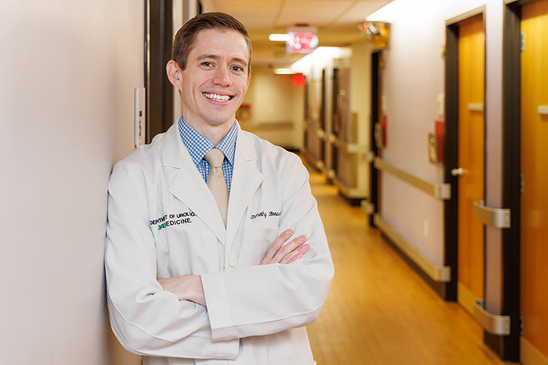
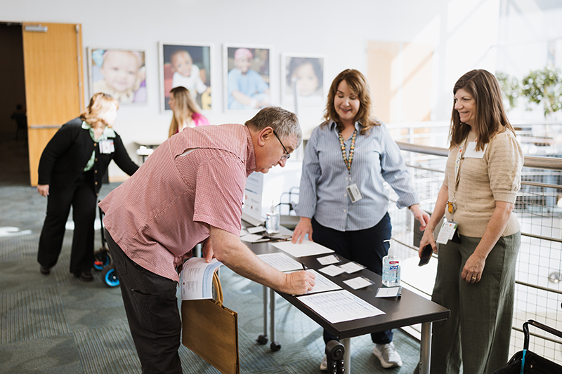
Gastroenterology

A high-tech approach to improving IBD treatment

When Children’s of Alabama associate scientist Babajide Ojo, MS, Ph.D., was completing his doctoral degree at Oklahoma State University, one of his peers had a child with Crohn’s disease. Ojo was so struck by the burden of the condition and its treatment on the child and his classmate—who eventually had to drop out of the program—that he decided his research must focus on inflammatory bowel disease (IBD), which includes Crohn’s and ulcerative colitis.

“I realized there were ways to manage the disease but no cure, so I thought it could be a great opportunity to contribute to this field and find better ways to treat it,” said Ojo, who’s also an assistant professor of pediatrics in the Division of Gastroenterology, Hepatology, and Nutrition at the University of Alabama at Birmingham (UAB).

Five years later—and a year after he arrived at Children’s—Ojo has already made an impact, using a newer, high-tech method to tease apart why IBD starts and what makes it flare in hopes of optimizing ways to control it. While most IBD treatments concentrate on calming the immune system, they don’t often lead to long-term remission, especially in children. So Ojo and his colleagues are looking at a different piece of the puzzle: the epithelium, or lining of the gut, which is important in IBD but often overlooked in research.

“The field right now solely focuses on the immune component of the disease,” he said. “But the data indicates that a lot of patients lose response to medications over time. For us, I think focusing on the epithelium may help us discover a kind of treatment that can elongate remission in these patients.”

Ojo is using a cutting-edge approach called patient biopsy-derived organoids—tiny 3D models grown from patient tissue—to watch how gut stem cells grow and become different kinds of cells that comprise the gut lining. By doing this, he hopes to understand how this process differs in people with IBD.

“Among the models we have, organoids may be one of the perfect ones to study the epithelium,” Ojo said, noting that few other pediatric centers use human-derived organoids for research. “Because each one represents the molecular features of each patient, it may be a way of developing personalized treatments.”

Based on his organoid research, Ojo and his colleagues published findings in December 2025 in Nature Communications suggesting that blocking a key fat-control protein helped epithelium cells in children with ulcerative colitis to burn energy more normally, reduce stress and calm inflammation. The results indicate that fat metabolism problems in colon cells are a major contributor to ulcerative colitis, potentially pointing toward new treatment approaches.

By identifying epithelial-specific “control points” central to IBD, Ojo hopes therapies can be developed that, if they don’t work for all patients, could be used specifically in pediatric patients.

“This may supplement some of the immunotherapies on the market to help us help them achieve long-term remission,” he said. “If we don’t improve how we treat patients, IBD is really a lifelong disease. We hope to make it much more manageable and reduce their constant visits to clinic, if not eliminate them totally.”

Endocrinology

The Future of Type 1 Diabetes Management

The future of type 1 diabetes management may involve more than just insulin. (Stock photo)

Think type 1 diabetes (T1D), and you think of insulin. You think incurable. You think forever. But the introduction of the first disease-modifying drug, the immunomodulator teplizumab, along with investigational approaches such as beta-cell preservation, gene editing and stem cell therapy, show that what we thought was the final word was really just the opening argument.

“For so long, we treated type 1 diabetes symptomatically with insulin alone, which is absolutely essential and life-saving,” Children’s of Alabama pediatric endocrinologist Ambika P. Ashraf, M.D., said. “But if we recognize it as an autoimmune disease, then we need to treat the immune process driving it, not just the high blood sugars. You don’t treat rheumatoid arthritis by just controlling inflammation alone,” she said.

To highlight where the field is at this important point in time, Ashraf co-edited a journal supplement, a special collection of research articles on T1D, in Frontiers in Endocrinology.

The supplement, published as a collection of 14 peer-reviewed articles, brings together researchers from around the world to address immune modulation, beta-cell preservation, remission, screening, gene editing, stem cell therapies and metabolic memory. The goal was not to champion one therapy, Ashraf said, but to show how the future of type 1 diabetes care will be built from complementary approaches to slow, stop or even prevent disease progression.

Two articles are authored by researchers at Children’s and the University of Alabama at Birmingham (UAB).

“When you look at all this work together as a collection, you start thinking about different ways to approach the management of type 1 diabetes,” she said.

The idea emerged while Ashraf and her colleagues were outlining a chapter on disease-modifying therapy for a pediatric diabetes textbook. “There was simply too much important work happening,” she said. “It became clear that disease-modifying approaches in type 1 diabetes deserved a deeper, more focused conversation.”

When Frontiers invited proposals for a themed issue, the timing was perfect.

“We knew this was it—bringing together the growing body of research that is shifting type 1 diabetes from a condition we only manage to one we may be able to change, delay or even prevent,” she said.

The supplement focuses on three questions: Can we slow or stop the immune destruction of beta cells? Can we preserve insulin production?  Can we delay or prevent the onset of symptomatic type 1 diabetes?”

“Remission is so important because if you stop autoimmunity, then you can get back to having normal function,” she said. At the same time, beta cell preservation is also key. “They go hand in hand.”

Another theme in the supplement is screening for antibodies that predict type 1 diabetes risk before symptoms appear, which opens the door to interventions when they are most beneficial. But universal screening is only practical if therapies are effective and accessible, Ashraf said. “If we had a medication that we could confidently say, ‘Take this, and it would reliably reduce your risk of developing T1D,’ then universal screening would be transformative,” she added.

Several articles in the collection explore therapies that support beta‑cell health without suppressing the immune system but by strengthening the cells themselves. These include medications like verapamil, experimental TXNIP‑targeting drugs and high‑dose GABA, all of which aim to help insulin‑producing cells survive longer.

Perhaps the most important message of the supplement is that there likely will not be a one-size-fits-all approach for people with T1D, but a mix of options based on the individual patient. As Ashraf and her co-editors wrote in an editorial in the issue: “The innovative concepts highlighted here will undoubtedly shape the future of diabetology and inspire further research into state-of-the-art, disease-modifying therapies for T1D.”

Endocrinology

Increasing Foot Exams in Kids With Type 2 Diabetes

When you think about Type 2 diabetes in kids, you likely think about blood sugar, insulin and diet. But what about feet?

For kids—and adults—with diabetes, feet can be the canary in the coal mine when it comes to microvascular damage from high glucose levels. In the feet, neuropathy increases the risk of foot ulcers and even amputation. In the eyes, it can lead to vision loss, and in the kidneys to chronic kidney disease.

“Young people with type 2 diabetes are very different from adults,” said Mary Margaret Barr, M.D., a pediatric endocrinology fellow at Children’s of Alabama. “Their disease is usually much more aggressive. It causes issues earlier, and it can be harder to control.”

Current American Diabetes Association guidelines call for comprehensive foot exams upon diagnosis of type 2 diabetes and annually thereafter.

Yet when Barr set out to see how well clinicians documented complete foot exams in medical charts at Children’s, a large gap emerged.

Her review came after the hospital transitioned to a new electronic health record (EHR) system. She found that after the transition, documented foot exams in pediatric patients with type 2 diabetes fell from about one in four visits to almost none. “Basically, one or two kids out of 30 or 40 had a documented foot exam,” she said.

It wasn’t that clinicians didn’t know the guidelines; it was just different in the new system.

Recognizing that reality, the team launched a formal quality improvement project—later named UndeFEETed—to understand what was getting in the way and how to fix it.

A Small Exam That Takes Time—and Intention

A proper diabetic foot exam is not a glance at a pair of sockless feet. It requires time, tools and attention.

It starts with a neurological exam to test nerve sensation in the foot. A monofilament (soft strand of nylon) is tapped gently on the foot and toes to assess if the patient can feel where it is tapping without looking, followed by tuning fork vibration tests, a pinprick test, or a tendon hammer test to assess ankle reflexes. The vascular assessment includes checking blood flow in the foot and ankle.

For children—especially those who are younger or developmentally delayed—this exam can be challenging. And in a busy clinic, where providers juggle multiple screening requirements, preventive exams that aren’t automated can quietly fall away.

“There’s a really big list of screenings that come along with diabetes visits,” Barr noted. “It does get overwhelming.” But most of these other screenings are incorporated into the daily clinic flow, with documentation rates averaging more than 80%.

Plus, there’s no reimbursement for foot exams despite the time required to properly do one. “That’s frustrating,” Barr said.

She’s not surprised that when she presents the UndeFEETed data at conferences with other endocrinologists around the country. “Everyone’s like: Yeah—no one does foot exams.”

But a documented foot exam is more than just paperwork: it’s a safeguard that problems won’t be missed.

“If a foot problem was found, it could be addressed early rather than later,” Barr said.

That matters because children diagnosed with type 2 diabetes today will live with the disease for decades. Large studies show that by their mid-20s many already show signs of permanent nerve damage, Barr said.

Her team identified four main barriers to documented foot exams: lack of standardized documentation, limited access to exam tools, time pressures, and uncertainty about how to record the exam in the new EHR.

Physicians, nurse practitioners and clinic nurses worked together to redesign workflows. A new diabetes note template embedded foot exam documentation directly into routine visits. Supplies were in all exam rooms. Nurses helped flag when exams were due.

The team resisted adding pop-up alerts in the new EHR, which also helped. Instead, they built seamless, non-intrusive reminders into the charting process.

The results were striking. Within months, documented foot exams rose from less than 5% to more than 20%. Once more providers adopted the new template, documentation climbed to 50%—double the original goal.

However, after the initial changes were implemented and the system was running for a while, that figure dropped to about 30%. So Barr and her team are continuing to investigate other options in the EHR to help. They are also working with the nurses to identify the best way to communicate with the clinicians they work with most often, whether it’s a sticker or a certain word to help them remember to conduct a foot exam.

Barr hopes the work eventually leads to tracking long-term outcomes, including whether earlier and more consistent screening reduces complications. For now, the focus remains on getting the fundamentals right. “Our biggest goal is to make sure that kids who are about to graduate from pediatric care and go into the adult world have foot exams on record,” she said.

Cardiology

Pulmonary Vein Stenosis: No Longer a Death Sentence

The pulmonary vein and artery system (Stock image)

Pulmonary vein stenosis (PVS) is a rare yet serious cardiovascular condition marked by progressive narrowing of the veins that return oxygenated blood from the lungs to the heart. Until recently, it was usually considered a death sentence. “There’s no two ways about it,” said Children’s of Alabama pediatric cardiologist Matt Clark, M.D. “It only gets worse once it starts.”

“When diagnosed, we would tell families there was nothing more to offer and to transition to end-of-life type care,” added pediatric interventional cardiologist Stephen Clark, M.D. “Until recently, we didn’t have medical therapies that could treat this. Surgical intervention was generally unsuccessful and sometimes accelerated the progression of the disease.”

Indeed, PVS has historically had grim outcomes, with mortality rates as high as 60% or more. Right ventricular pressure elevation—a marker of pulmonary hypertension (PH)—is strongly associated with mortality.

Now, there is hope.

“I think the biggest thing to come along was more data from studies and centers showing if you change the mindset and consider it as a possibly treatable condition, you change outcomes,” Matt Clark said.

That shift in mindset has driven a transformation in how PVS is diagnosed, monitored and treated. Today, children who would not have survived infancy are living into early childhood—and in some cases beyond—because of earlier recognition, aggressive catheter-based interventions and multidisciplinary care.

Earlier Detection, Closer Surveillance

One of the most important changes in PVS care has been earlier diagnosis, Matt Clark said, by recognizing high-risk populations: neonates with chronic lung disease; prolonged ventilation; those with congenital heart disease; and/or those with pulmonary hypertension (PH). The earlier the diagnosis, the more likely interventions are to be effective.

This surveillance-driven approach means clinicians are often identifying milder disease earlier—with regular echocardiograms, CT scans and diagnostic catheterization—and intervening at the first signs of progression.

PVS often exists in the setting of comorbid conditions such as PH, congenital heart disease or complications of prematurity, requiring close coordination between specialists. Thus, the pulmonary vein stenosis team in the Children’s of Alabama Heart Center has brought together a small group of pediatric cardiologists, allowing for shared memory and standardized decision-making.

“We don’t have to spend time reinventing the wheel,” Matt Clark said. “We can collectively remember all our patients” and which approaches approach worked best.

Catheter-Based Intervention as a Strategy, Not a Last Resort

Treatment for PVS has also evolved. Intervention includes balloon angioplasty to widen narrowed vessels or stent placement to hold them open. The stents are coated with sirolimus, a drug designed to prevent restenosis. However, rather than viewing catheter-based intervention to unblock the vessel as a one-time attempt to rescue a sick child, it is now viewed as a repeated, planned strategy.

“Every time you do an intervention,” Matt Clark said, “you’re signing that kid up for another cath at some interval.” That’s because the baby’s heart and lungs keep growing, requiring additional interventions. Multicenter analyses show that repeat catheterizations are associated with improved survival, reflecting a shift away from passive observation to active disease control.

In some children, repeated dilations and stent expansions over time allow pulmonary veins to reach near-adult size within the first few years of life.

Medical Therapy to Slow Disease Progression

Procedural care is now complemented by medical therapy aimed at the underlying biology of PVS.

Because the disease involves aggressive cellular proliferation within the vessel wall, antiproliferative medications such as sirolimus have become an important adjunct to intervention.

Observational studies suggest that children receiving systemic sirolimus have significantly improved survival compared with historical controls. In one widely cited cohort, four-year survival reached 100% in treated patients compared with approximately 45% in those not receiving therapy.

Sirolimus has also been associated with slower in-stent restenosis and longer intervals between interventions. While randomized trials are still lacking, these findings have helped legitimize systemic therapy as part of standard care at experienced centers.

“We’re probably doing more sirolimus-type therapies now than we used to,” Matt Clark said.

Survival Looks Different Now

Even with advances, PVS remains unpredictable. Not every child can be saved. But survival is no longer measured only in weeks or months.

Matt Clark described children who once would have died in the hospital now going home, growing and returning for planned interventions.

“One of the earlier ones was pretty impactful,” he said, recalling a child who lived long enough to spend two Christmases at home. “That was enough to make us keep going and keep pushing.”

“We know we’re helping,” he said. “So we’re more likely to intervene, to follow closely and to keep trying.”

Cardiology

Building a Team Around Pulmonary Hypertension

The Children’s PH team (L to R): Ahmad Khalil, Frank Bennett Pearce, Jodie Kanaday, Kevin Wall and Matthew Clark.

Pulmonary hypertension (PH) is a heterogenous condition stemming from numerous underlying causes, including extreme prematurity, congenital heart disease and systemic diseases like lupus. It causes dangerous increases in blood pressure in the lungs, straining the heart’s pumping ability and potentially leading to heart failure.

Care for children with PH has typically existed across silos: cardiology, pulmonology, neonatology and intensive care, said Frank Bennett Pearce, M.D., a pediatric cardiologist at Children’s of Alabama. Each discipline did its part. But clinicians increasingly recognized that the complexity of the condition and the fragility of the patients required greater coordination.

Today, a formal PH team of Pearce, pediatric cardiac critical care specialists Ahmad Khalil, M.D., and Matthew Clark, M.D., and pediatric cardiology fellow Kevin Wall, M.D., together with pediatric cardiology nurse specialist Jodie Kanaday, RN, round weekly on patients with PH to determine the best course of action for this rare but complex condition.

“We wanted to improve communication by having us all together saying the same thing to the consultant teams and to the families,” Pearce said.

That consistency matters, especially when care unfolds over weeks or months.

From a cardiology standpoint, PH has always been part of the landscape, Pearce said. “A lot of the treatment and diagnostic procedures like catheterizations and echocardiograms come through cardiology anyway,” he said. “So we end up being the treating doctors in lots of cases, or at least consultants.”

At the same time, many of the sickest patients are in neonatal and pediatric intensive care units and managed by critical care specialists.

PH is typically treated with medications like pulmonary vasodilators regardless of cause, but timing and diagnosis matter. That’s why it’s so important to have a precise anatomic diagnosis before starting medication, Pearce said. In babies with bronchopulmonary dysplasia, for instance, PH may be driven by acquired pulmonary vein stenosis—a condition that requires catheter-based or surgical intervention before medication.

Previously, decisions like these might have been made in parallel by different services. Now, they are made together.  

The team manages about a dozen inpatient pulmonary hypertension cases each month on inpatients at Children’s and the University of Alabama Birmingham (UAB). But inpatient care is only part of the story.

PH does not end at discharge. Medication management, insurance approvals, symptom monitoring and urgent questions follow families home. That’s where Kanaday, who is the Pulmonary Hypertension Clinic care coordinator, shines.

“We literally couldn’t do it without her,” Pearce said. She allows the team to see more patients, stay more organized and keep up with the paperwork/regulatory side of things in the PH world, he said.

Kanaday sees herself as a conduit to the physician. “Our patients know they can contact me directly for questions about medications, symptoms or side effects. I’m able to pass those concerns along quickly, which usually means they get help faster than they otherwise would.”

Her role, she said, is “making sure the doctors have all the information that they need to best take care of the patients.”

While the team is not formally tracking outcomes yet, Pearce says he’s seen a difference. “The patients are staying on their medications more consistently. The doses are more consistent. Some of these medications require a lot of paperwork—that’s getting handled quicker.”

In addition, the improved coordination means patients get to the cath lab sooner for pulmonary vein interventions. The team is also closely tied to newer catheter-based procedures, such as closing a patent ductus arteriosus or atrial sepatal defects in extremely small infants.

“I think it’s really been an asset to our cardiology team in general, taking this burden off of the general cardiologists and putting these patients with a provider who readily understands the disease process, available treatment options and possible complications. I think it makes our consult service much more efficient,” Kanaday said. “It has really made an impact on the patients, their continuity of care as an inpatient, and their follow-up in the outpatient clinic. I feel like we’re really making a difference and giving these kids the best chance to have positive outcomes long term.”

Urology

In New Role, Hopson Working to Address Sexuality in Patients With Spina Bifida

Betsy Hopson, Ph.D., MSHA, recently moved into a new role within the Division of Pediatric Urology.

Betsy Hopson, Ph.D., MSHA, describes her philosophy in one word: listen.

“Early in my career, I adopted this principle that if I heard the same story from two or more patients then it was either a research question or a quality improvement opportunity,” she said.

That mindset has shaped nearly two decades of work at Children’s of Alabama and the University of Alabama at Birmingham (UAB) and now underpins her new role as an assistant professor and health scientist in the Division of Pediatric Urology. There, she will direct a clinic dedicated to helping children with congenital urologic conditions transition to the adult health care setting.

Hopson began her career at Children’s in 2006 as coordinator of the Spina Bifida Program in the Division of Pediatric Neurosurgery. Yet she spent nearly as much time interacting with urology, she said, which plays a central role in bladder management for patients with the disease.

Her early work focused on helping patients make the difficult transition from pediatric to adult care. After realizing that young adults with spina bifida were aging out of pediatric clinics without clear adult pathways, she returned to school to earn a master’s degree in health care administration and then built an internationally recognized transition model.

It was listening to her patients, however, that led to the next chapter in her life.

In one case, a 15-year-old adolescent asked her about her research. When she told him it was sexual and reproductive health, “He whispered, ‘Betsy, I can’t do that, can I?”’

“Can’t do what, buddy?” she asked. “Date or have sex,” he answered. “That’s not for me, is it?”

Or the newly engaged, college-educated young woman with spina bifida who burst into tears when Hopson started discussing birth control with her. “You mean I can get pregnant?” she said. “I had no idea.”

“If this young woman who is very educated had no idea,” Hopson said, “what is this like for the rest of the population?”

And a light bulb went off. “We’re telling them we want them to be independent, that we want them to take care of themselves, but we’re not giving them any carrot or showing them what’s possible and helping them paint that picture of what adult life could look like.”

Her “eureka” moment led her back to school to obtain her Ph.D. in Rehabilitation Science and a certificate in Mixed Methods Research. Her goal was to understand gaps in sexual health education for people with congenital diseases like spina bifida. What she uncovered was far more troubling.

“Because these patients are prescribed catheterization for bladder management early in life,” she said, “they’re taught the technical skills of catheterization but not taught about personal boundaries and appropriate touches.”

Her research found that 46% of adults with spina bifida reported a history of sexual abuse. For her dissertation, she validated a clinical screening tool to identify abuse risk and gaps in sexual health knowledge.

In her new role with the urology team, she sees her goal as twofold: “One is to help support normal development,” she said. “I want to normalize sexual and reproductive health conversations so patients can see what’s possible and give them space to imagine a full adult life.”

The other involves educating patients, parents and clinicians about the increased risks vulnerable patients face and developing tools for clinicians and families to identify and talk openly about those risks.

It’s important, she noted, to bring the parents into the discussion. “When you bring [sexuality] up in the clinical setting in front of their child, they might be initially hesitant.” Her solution is the same approach that has guided her career: listen first. That means holding focus groups with patients and parents to shape new curricula and ensure the content reflects lived experience rather than clinician assumptions.

“If there’s one thing my career has taught me,” she said, “it’s to never stop learning and never stop looking for ways to make a difference.”

Cardiology, Inside Pediatrics

Lau shares vision as new Physician-in-Chief

Yung Lau, M.D. was named physician-in-chief at Children’s of Alabama and chair of the UAB Dept. of Pediatrics in March 2025.

After serving as interim chair for five months, Yung Lau, M.D., was officially named chair of the University of Alabama at Birmingham (UAB) Department of Pediatrics and physician-in-chief at Children’s of Alabama in March 2025. The only real change, Lau said, was that he could now formally begin planning for the future. His vision for the department is expansive—centered on collaboration and faculty support. But he believes the path to those big goals lies in the small things everyone does every day.

Lau stepped into the interim chair role in November 2024, following the announcement that then-chair Mitch Cohen, M.D., would be departing at year’s end to join Stanford. “Dr. Cohen led for a decade and helped build this department into a strong and vibrant group,” Lau said. “It has consistently thrived, and I’ve considered it a privilege to be a part of the department as a faculty member for over three decades. Now, it’s an awesome responsibility to carry on this tradition of excellence.”

The role requires close collaboration between two major institutions: Children’s of Alabama and the University of Alabama at Birmingham. With more than 30 years of experience across both organizations—including in leadership roles—Lau understands their individual missions and how they intersect.

That understanding came into sharp focus in 2007, when Lau led the UAB group in a major collaborative effort between the two institutions. At the time, UAB housed the pediatric cardiac program. But as Children’s leaders planned to build a new hospital tower, they wanted to bring the program under their roof. Over the next five years, Lau played a significant role in bringing physicians and other clinical staff to assist in the design of the cardiac intensive care units, operating rooms, catheterization labs, step-down units and cardiovascular perioperative areas. He then worked closely with Children’s leadership on staffing and operations planning. On October 14, 2012, the program moved into the new building—seamlessly.

The success of the move laid the foundation for a quantum leap in the ability of the Pediatric and Congenital Heart Center of Alabama to provide state-of-the-art care. Today, the program consistently ranks among the top performing pediatric and congenital heart centers in the nation by numerous metrics, and the Society of Thoracic Surgery has classified the program as an overperforming center—one of 12 in the country. For Lau, who served as the division director of pediatric cardiology from 2012 until his appointment as chair in 2025, the experience left a lasting impression about what’s possible when Children’s and UAB work together.

“The opening of the Heart Center marked one of the most satisfying periods of my career,” he said. As he steps into his new role, he hopes to lead more collaborative efforts with similarly meaningful impact.

The Heart Center’s success is a powerful example of the synergy between Children’s and UAB—a synergy Lau believes can grow even stronger. “What I’ve seen is a real willingness among leadership across both institutions to reduce barriers and connect silos,” he said.

Lau has outlined three key priorities to strengthen that collaboration: maximizing current resources in clinical care, education and research; strategically recruiting and developing faculty; and building a resilient foundation of financial stability and physician well-being.

Education and research are crucial parts of his strategy, and UAB and Children’s have a history of successful collaboration on both. From an education perspective, Children’s serves as the teaching hospital for the UAB pediatric medicine, surgery, psychiatry, research and residency programs. “There’s this deep core of understanding between Children’s and the department that we are really training the future physicians for the state,” Lau said. And that’s a crucial role in a state that, Lau says, needs more physicians and pediatricians. “Part of our duty here is in our obligation to do that,” he added.

On the research side, the two institutions work together to “advance knowledge for the children of Alabama and beyond for the future,” Lau said. This benefits both entities, sometimes leads to advancements and breakthroughs that influence the broader world of medicine, and enhances the reputation of both.

“When we collaborate more extensively and continue to strengthen those ties of collaboration, two important things happen,” he said. “First, children receive better care, now and in the future. Second, our faculty experience significantly greater job satisfaction.”

Faculty support is another central pillar of Lau’s vision. Since becoming chair, he has met with many faculty members—some he’s long known, others he’s come to know better through these conversations. What stands out most, he says, is their passion and the profound impact they have on children’s lives. His goal is to listen, support and help them succeed.

Lau also acknowledges the tension between moral obligation and financial reality. “That’s just medicine in America today,” he said. But he’s confident the department can thrive within that framework.

“I think both institutions understand that we need to maximize our resources—our people and infrastructure—to provide the best possible care, to train the next generation of pediatricians, and to innovate through research,” he said.

Though there are multiple facets to Lau’s vision, everything is focused around the patient.

“The patients in front of us are the cornerstone of everything we do,” he said. “And while our goals may be big, the real progress happens in the small steps we take every day.”

“Yes, having a goal is important,” he continued. “But sometimes if we focus only on the goal, we risk losing sight of what’s happening in the moment—and that can distort the work being done on the ground. Sometimes the things that matter most get sidelined in the name of progress.”

With a strong focus on the patient—and through strong collaboration and faculty support—Lau believes UAB and Children’s will continue to deliver exceptional care to every child they serve.

Neurology & Neurosurgery

An endoscopic approach for skull base conditions

Dr. Jessica Grayson (left) and Dr. James Johnston perform an endoscopic procedure on a patient at Children’s of Alabama.

A growing number of children with complex skull base conditions can now be treated with minimally invasive surgery at Children’s of Alabama thanks to a collaboration between pediatric neurosurgeon James M. Johnston, M.D. and otolaryngologist Jessica Grayson, M.D. Together, they lead an integrated pediatric skull base surgery program that offers endoscopic procedures for conditions such as skull base tumors, traumatic injuries, complex pituitary lesions, and congenital abnormalities such as encephaloceles—in which brain tissue protrudes through an opening in the skull.

“Endoscopic approaches have been part of pediatric neurosurgery here for years for things like hydrocephalus or intraventricular tumors,” said Johnston, director of the Division of Pediatric Neurosurgery at Children’s and the University of Alabama at Birmingham (UAB). “What’s new and exciting is how we’ve expanded endonasal skull base surgery through this collaboration.”

The procedure involves threading a tiny camera and instruments through the patient’s nasal passages to reach the brain. “That means smaller incisions, less blood loss and a much shorter recovery time,” said Grayson—one of the few clinicians in the country who is fellowship trained in rhinology and skull base surgery for both adults and children, with extensive expertise in endoscopic endonasal surgery.

After tumor removal, Grayson works to patch any small holes created between the brain and the nose. This is one of the most critical aspects after the removal—if the small holes aren’t properly sealed, cerebral spinal fluid could leak out into the nose, leading to a high risk of infection. Grayson typically uses a nasoseptal flap to close any openings. She peels a small piece of the mucosa covering the nasal septum while maintaining its blood supply, then flips it over to cover any holes created during surgery.

The program is multidisciplinary, involving ENT, neurosurgery and occasionally plastic surgery. The team-based model also allows for comprehensive case review and planning. “We often consult with our adult colleagues at UAB when a case is really complex,” Johnston said. “It’s like having a built-in tumor board.”

Offering this type of approach for children is another way the program is unique—this method typically has been reserved for adults. And “the technical aspects are different from adult cases,” given their smaller anatomy and less-developed sinuses, Johnston noted. “But with collaboration, it’s absolutely feasible. We’ve even done this in infants as young as a few months old.”

The first collaboration—a case of congenital encephalocele in which the protruding tissue was initially mistaken for adenoid tissue—highlighted the potential of combining expertise. “That was the moment we realized we could safely and effectively treat these cases together using a minimally invasive endoscopic approach,” Grayson said.

Nationally, this type of program is rare. “In many places, kids are sent to adult hospitals for these procedures,” Grayson said. “Here, they can stay in a pediatric environment with pediatric anesthesiologists, nurses and postoperative care, which is crucial for safety and comfort.”

Last year, the team did about 40 cases, and the number of referrals is growing as more clinicians become aware of what’s possible. “We’re seeing more cases from outside hospitals,” Johnston said. “And we’re better at recognizing which patients are good candidates.”

Gastroenterology

New technology eases management of liver disease

Children’s of Alabama is using FibroScan to help patients with liver disease. (Stock photo)

With obesity in children steadily rising, more young patients are coming to Children’s of Alabama with a form of fatty liver disease that can greatly imperil their health. But determining the progression of liver disease can be a thorny process. To smooth that path, Children’s recently invested in an increasingly popular technology called FibroScan, helping University of Alabama at Birmingham (UAB) pediatric physicians to deftly and comprehensively manage children’s care.

Using a technique known as transient elastography, FibroScan was the first FDA-approved device of its kind and is considered an aid to managing liver disease. Quick, noninvasive and painless, it uses an enhanced form of ultrasound to send vibrations into the liver to measure its stiffness, which typically indicates fibrosis or scarring. “The more quickly the wave passes through the liver, the more stiff the liver is,” Children’s transplant hepatologist David Willcutts, M.D., explained.

By assessing the severity of scarring—and the potential for cirrhosis—FibroScan can help diagnose or monitor the progression of various liver conditions. These range from less-common cystic fibrosis-associated liver disease to more-prevalent autoimmune liver diseases and metabolic dysfunction-associated steatotic liver disease (MASLD). The latter—which can also result from genetic predisposition—essentially makes the liver unable to process the high amounts of extra calories a person is consuming, spurring inflammation.

David Willcutts, M.D.

About one-third of the patients in Children’s Hepatology Clinic, which serves about 500 ongoing patients each year, have suspected or confirmed fatty liver disease.

“We will be using this for almost every patient with confirmed fatty liver disease, so we can measure the baseline stiffness of the liver when they first see us,” said Willcutts, who’s also an assistant professor of pediatrics at UAB. “The machine also provides a CAP (controlled attenuation parameter) score as a surrogate of fat content of the liver, which is useful for the growing numbers of adults—and unfortunately, children—in our country with fatty liver disease. It’s one of the rising conditions leading to adult liver transplants.”

FibroScan is a welcome alternative to invasive liver biopsies and other forms of elastography that require a separate radiology appointment. A FibroScan exam takes just minutes, offering little disruption for young patients and faster treatment decisions for physicians. The new equipment arrived in the summer of 2025.  

“One of the big selling points of this technology is it makes the patient experience much easier because it can be done within a clinic visit and will save them a visit with radiology, which involves a separate appointment elsewhere in the hospital or even at another Children’s facility,” Willcutts said. “It’s a one-stop kind of assessment.”

By keeping close tabs on a patient’s liver stiffness, FibroScan offers Children’s specialists the ability to understand “how much runway we have before we need to do potentially invasive assessments and other therapies,” Willcutts said.

While the goal is always to avert lasting damage to the liver, the presence of cirrhosis is generally thought to be irreversible. FibroScan can help doctors pinpoint “how close we’re getting to that and if the patient needs a biopsy—or a repeat biopsy—to evaluate scarring at the microscopic level and make sure we’re not missing something before it’s too late to act upon it,” he explained.

FibroScan results can also help physicians tailor treatments to patients’ precise stage of liver damage, including certain medications that can be tricky for the liver to process.

“Children’s is a referral center for pediatric liver disease in Alabama because we’re the only liver transplant center in the state,” Willcutts said. “Being able to offer FibroScan helps us elevate our level of care and offer smoother visits and a convenient assessment of liver disease that we didn’t have before.”

Nephrology

Leading-Edge Technology May Change Kidney Transplant Monitoring and Help Other Specialties

In the UAB Spatial Core Lab, researchers are using spatial transcriptomics to examine specific regions within tissue samples.

A breakthrough technology that allows doctors to study the precise regions of a kidney transplant that are involved in rejection could transform how doctors learn about kidney transplant biology, potentially leading to new diagnostic tests or treatments for children.

The technology, called spatial transcriptomics, is a leading-edge technique that lets researchers see which genes or proteins are active in very specific regions of tissue samples, such as those from a kidney biopsy. Unlike traditional methods that study tissue samples as a whole without regard for the location of important signals from the tissue, this technique allows researchers to examine custom-shaped regions of interest containing just a few cells in their natural environment.

Think of it as having a detailed map that shows not just what’s happening in a city, but precisely in which neighborhoods each activity occurs.

“There’s been decades now of data showing that gene expression patterns coming from a transplant are a little bit more sensitive for problems coming from a kidney transplant,” said Michael Seifert, M.D., director of the University of Alabama at Birmingham (UAB) Spatial Core and medical director of pediatric renal transplantation at Children’s of Alabama. “The problem is that we’ve never exactly known where those signals are coming from. Are they coming from cells in the kidney that we care about or are they coming from cells in the kidney that may not be as relevant?”

For instance, signals from immune system cells would be extremely relevant, he said, but could be distinct from those coming from the endothelial cells lining blood vessels.

With this technique, “we can look at a picture of a kidney biopsy on our instrument screen and take your mouse and draw a shape around it, and it will profile everything in that shape while ignoring everything else around it,” he said. It can even profile a certain cell type within the shape.

The Spatial Core team (from left): Pooja Nagaraj, MS, CCRP, Michael Seifert, M.D., Miguel Melendez-Ferro, Ph.D.

The technology itself isn’t destined for routine clinical use, Seifert said. “I can’t foresee a scenario where I would do a biopsy and then use spatial transcriptomics to make a diagnosis, because it’s a very labor-intensive, time-intensive and cost-intensive technique.”

Instead, he said, “my hope is that this will allow us to have a deeper understanding of the processes involved in transplants doing well but also transplants doing poorly. That will help us design better management programs, whether that’s using existing medicines in different ways or designing new medicines that can be more targeted and more effective than what we currently have available.”

Understanding exactly which parts of the kidney are affected by rejection also opens the door to personalized transplant care.

“Every cell in the kidney behaves differently depending on where it sits,” Seifert said. “This technology lets us uncover the heterogeneity—that is, the differences—within the tissue,” including if the problem lies in the blood vessels or the tubules or the parts of the kidney that generates urine. “I hope that’ll allow us to understand the signals that vary from person to person so we can really apply that more personalized technique.”

Thus, rather than treating all kidney transplant patients the same way, doctors could tailor anti-rejection treatments based on what is happening in an individual child’s kidney. This would, however, require advances in the spatial transcriptomics technology to make it faster and less expensive.

Spatial biology is not limited to the study of kidney transplant diseases. Seifert and his team in the UAB Spatial Core are working with specialists in other disciplines throughout Children’s and UAB, including ophthalmology, oncology and pulmonology. “We’re open to collaborating with any investigator with a good question that spatial biology can answer,” he said.

In fact, he sees spatial biology as an important technique for understanding all diseases in children. “I think what’s come out of this is an appreciation that the spatial context is incredibly important in so many of the diseases that we study.”