Browsing Category

Neurology & Neurosurgery

Neurology & Neurosurgery

Deep brain stimulation for progressive dystonia

In 2023, Children’s of Alabama performed deep brain stimulation on progressive dystonia patients for the first time.

Progressive dystonia disorders, characterized by changes in movement patterns, can profoundly impact a child’s quality of life. In adults, such disorders are routinely treated with a procedure called deep brain stimulation (DBS). However, this intervention is less commonly used in pediatric populations.

In 2023, Curtis Rozzelle, M.D., and Emily Gantz, M.D., performed Children’s of Alabama’s first DBS procedures for progressive dystonia patients. This innovative therapy has shown promising results in several pediatric patients with limited treatment options.

Curtis Rozzelle, M.D.

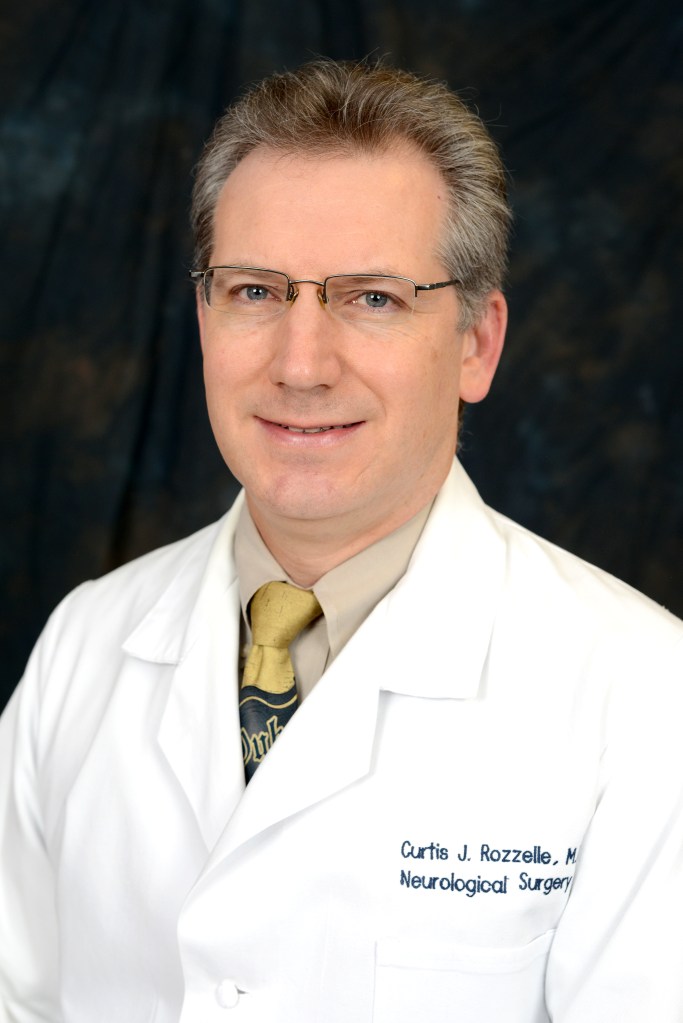
“We’re excited that this innovative procedure is now transferring over to our pediatric patients,” Rozzelle, a pediatric neurosurgeon at Children’s, said. “Kids with progressive dystonia seem to do particularly well, while those with other types of movement disorders may have mixed results.”

Studies suggest that pediatric patients with progressive dystonia respond well to deep brain stimulation, especially after failing conventional medications. The decision to apply DBS in pediatric cases stems from the specific needs of young patients and advancements in the field.

“Until Dr. Gantz arrived at the University of Alabama at Birmingham, we didn’t have a movement disorder neurologist here who also had training and experience with deep brain stimulation,” Rozzelle said. “When she arrived, Dr. Gantz opened the door for us to be able to perform the technical aspects of the surgical procedure. Now, we’ve done several.”

Not every child is an ideal candidate for DBS. The decision to initiate DBS is patient-specific, based on the severity of symptoms and the inadequacy of other treatments. A careful evaluation of each patient’s unique situation, including factors such as genetic mutations and the progression of the disease, must be completed before offering DBS surgery.

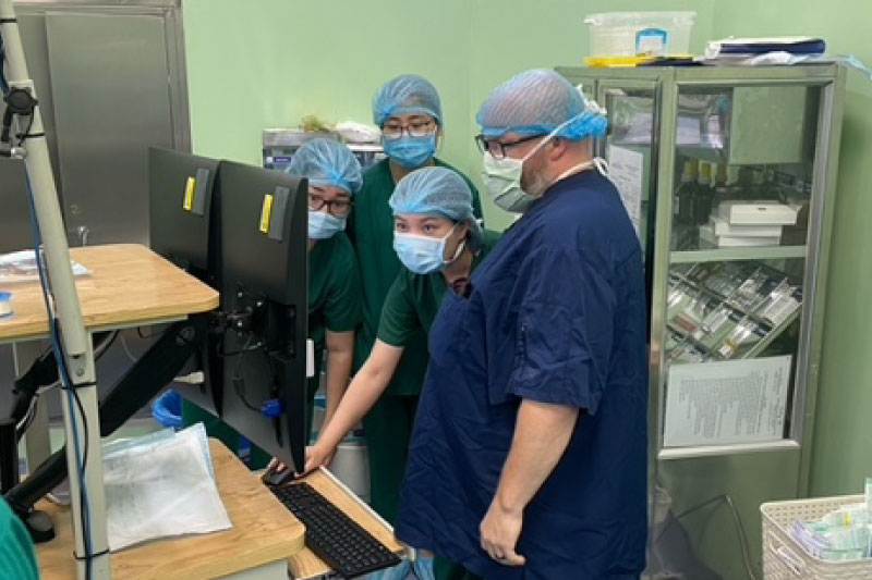
Each deep brain stimulation procedure at Children’s uses stereotactic surgical techniques and the ClearPoint targeting system. This system, employed in an MRI scanner, ensures safe, precise electrode placement in the globus pallidus interna (GPI), a key target for treating progressive dystonia.

Emily Gantz, M.D.

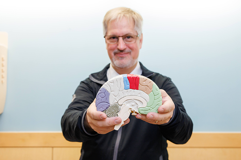
“When we implant the electrode into a specific region of the brain, we can either edit the input throughout the stimulation, or we can take it completely away,” Gantz said. “Think of it as a series of relay circuits in the brain. If someone has dystonia, one of those relay circuits isn’t working properly. By putting in the stimulator and applying an electrical current intermittently, we can suppress the abnormal brain activity.

“The stimulator stays in for life, so the procedure doesn’t need to be repeated,” she continued. “Occasionally, we’ll have to change the device’s battery, but they’re rechargeable and designed to last for up to 20 years.”

After the initial procedure, patients return to see Gantz to have the device programmed. “I set the programming on their stimulator so they can make slight adjustments at home. It can take a little while for the device to be effective; we usually leave it alone for a few months and then reevaluate,” Gantz said. “We have guidelines for which settings will most likely help, and we start there. We’re looking to ensure we don’t get side effects, such as visual disturbances or muscle pulling, more than anything.”

The pediatric patients who have begun DBS for progressive dystonia at Children’s are responding well to the new treatment. “I’m really excited about DBS and its future as a treatment in pediatric neurology, specifically movement disorders,” Gantz said. “It may eventually come into play in other treatment areas, and I’m glad the door is open to us here. I think there will be many more patients who will benefit from it.”

Neurology & Neurosurgery

A new procedure for epilepsy patients in Vietnam

Children’s of Alabama director of neurophysiology Trei King with a Vietnamese EEG team during a trip to Vietnam in September 2023. (Submitted photo)

With the waning of the COVID-19 pandemic, a team of neurosurgeons from Children’s of Alabama, Johns Hopkins All Children’s Hospital and Nationwide Children’s Hospital in Columbus, Ohio, were finally able to fly the 9,000 miles back to Vietnam in 2023 to continue training surgeons on surgical techniques to manage drug-resistant epilepsy.

Children’s of Alabama’s relationship with Vietnamese neurosurgeons began in 2013 with an initial visit to a team in Ho Chi Minh City. Until the pandemic hit, the team, including pediatric neurosurgeon Brandon Rocque, M.D., pediatric epilepsy surgery director Pongkiat Kankirawatana, M.D., and director of neurophysiology Trei King, BA, R.EEG.T, CNIM, visited annually to provide hands-on training at hospitals in Hanoi and Ho Chi Minh City.

Their efforts are desperately needed in a country with just two adult and two pediatric neurosurgery training programs for its 95 million people and only six pediatric neurosurgeons serving a population of more than 50 million in the northern part of the country.

“Vietnam did a very good job of managing COVID, with an extremely low per capita death rate,” Rocque said. Nonetheless, there were significant disruptions to medical care and training during lockdowns.

On their return trip to Vietnam in September 2023, the team assisted surgeons in Ho Chi Minh City with epilepsy resection surgeries. Since the Children’s team left, the local surgeons have completed at least two of these procedures on their own, albeit with some long-distance help from the Children’s surgeons. “They called us in the middle of the night, and we helped them troubleshoot the equipment a bit for the epilepsy monitoring,” Rocque said. 

On the same trip, at the National Children’s Hospital in Hanoi, the team performed the country’s first subdural grid electrode implantation, a procedure designed to pinpoint where seizures are occurring. “Everything went really well,” Rocque said. “We monitored the patients for a couple of days and were able to clearly localize where their seizures were.” Then, they removed the electrodes and performed the resection.

The procedure had never been performed in Vietnam because of concerns about infection from the temporary electrodes and the need to keep patients heavily sedated. However, those concerns were overcome when the hospital adopted international standards for the procedure.

The grid implantation, performed in two pediatric patients, received national media coverage, triggering requests from families throughout the country. “It opens up the possibility of many more patients getting treated,” Rocque said.

The team also visited the National Cancer Hospital in Hanoi to assist with an established program using selective dorsal rhizotomy to reduce spasticity in the legs from cerebral palsy. They helped evaluate patients, assisted with surgery and participated in a symposium on the procedure attended by more than 50 physicians throughout Vietnam.

The team also assisted the Vietnamese neurosurgeons in performing extraoperative video-electrocorticogram monitoring.

Neurology & Neurosurgery

Global Alliance Co-Founded by Children’s Neurosurgeon Affecting Change in Spina Bifida Prevention Effort

Spina bifida is the most frequently occurring permanently disabling birth defect to affect the nervous system.

Jeffrey Blount, M.D., MPH, knows the struggles of patients with spina bifida (SB). He and his colleagues in the Division of Pediatric Neurosurgery at Children’s of Alabama and the University of Alabama at Birmingham (UAB) have seen them firsthand—hydrocephalus, lower extremity paralysis, sleep apnea, pressure sores, variable incontinence, and the frequent need for multiple surgeries. Other doctors providing SB care see urologic, musculoskeletal, orthotic and ambulatory problems. A few years ago, the desire to address these issues led Blount to a big idea—one that would help not only his patients, but others around the world. In 2019, he co-founded the Global Alliance for the Prevention of Spina Bifida, or GAPSBiF, an organization dedicated to increasing awareness and advocating for the prevention of SB through large-scale food fortification with folic acid (FA). It’s already affecting change.

Blount is the medical director of the Pediatric Spina Bifida Clinic at Children’s of Alabama—one of the largest clinics of its kind in North America, following about 450 children. The medical professionals in the clinic work with those at the Adult Spina Bifida Clinic at UAB, which follows about 250 adults. In founding GAPSBiF, Blount partnered with Gail Rosseau, M.D., an international leader in global neurosurgery; Adrian Caceres, M.D., a Costa Rican neurosurgeon who accomplished widespread fortification of FA in Costa Rica; and Colombian neurosurgeon Kemel A. Ghotme, M.D., Ph.D., who had just completed a Ph.D. in Global Health Policy with a focus on FA fortification. One of the GAPSBiF’s major strategies for preventing SB was working with other neurosurgical and nutrition directed organizations in putting together a resolution that called upon all World Health Assembly (WHA) member states to embrace micronutrient fortification including FA to prevent SB. Resolution 76.19 was introduced by the Colombian government and 37 other member states and went through a rigorous process of vetting. In May, the WHA adopted it.

“This has real potential to favorably and fundamentally impact the global prevalence of SB and other micronutrient dependent diseases,” Blount said. “It is an essential step toward overcoming the stalled progress on the prevention of spina bifida.”

Spina bifida and folic acid

SB is the most frequently occurring permanently disabling birth defect to affect the nervous system. It results from the spine’s failure to close properly during the first month of pregnancy. The cause of SB is not fully understood, but it is thought to be associated with both genetic and environmental factors. The most important environmental factor is maternal intake of dietary FA, a B vitamin that is critically important in development and has long been known to reduce the risk of neural tube defects (NTDs), such as SB.

Nutritional shortage of FA in women of childbearing age is the most important contributor to SB prevalence worldwide. Many women supplement FA in their diet by taking 400 micrograms of FA while pregnant. But, in some cases, that’s not soon enough. “This problem of spina bifida occurs so early on in development that it has already occurred before most women even realize they’re pregnant,” Blount said. “So, it’s not like they can realize they’re pregnant, change their nutritional strategy and put up an effective barrier for this problem. Once they realize they’re pregnant, if they have the problem, it’s already occurred.” Fortifying widely consumed foods such as corn, grain or rice is more effective, which is why GAPSBiF works so hard to promote this strategy.

Evidence that fortification helps

In the U.S., mandatory fortification of enriched cereal grain products with FA was authorized in 1996 and fully implemented in 1998. Here, NTDs, including SB, affect approximately seven out of every 10,000 births. The rates in other regions that fortify are similar. In regions that don’t fortify, NTDs affect up to 150 births per 10,000.

But some countries—even advanced Western European nations—still are not practicing fortification, and, in many cases, are focused more on detection. But that approach can be problematic, Blount says. “Some places are very aggressive at terminating those pregnancies, which of course is a very difficult, very challenging, whole approach to problems. But it’s surprisingly widespread.”

GAPSBiF’s approach is centered around prevention. “Let’s keep these little children from getting this terribly difficult disease,” Blount said, “because it’s lifelong.”

The role of GAPSBiF

When Blount and his colleagues were forming GAPSBiF, they spoke with neurosurgeons from around the world. Even in North America, where fortification is already commonplace, SB takes an exhausting toll on patients, families, the health care system and the neurosurgical infrastructure. In many other countries, it’s much worse—due not only to the lack of fortification, but also because there are far fewer neurosurgeons per person. “A big part of their life and their world is taken up caring for these children,” Blount said. “And it prevents them from being able to do other things, such as taking care of people with strokes, taking care of people with trauma, things like that. So, it overloads an already challenged workforce.

“We saw this, we came together as a group, and we said, ‘Neurosurgery sees this. Neurosurgery knows this disease. We have a front-row seat to all these problems. So, why don’t we try to organize in such a way that we work with other agencies to try and attain this goal of universal fortification?’ ” Blount said.

“We know that if we can get folic acid into population food supplies, that up to 90% [of the SB cases worldwide] can be profoundly reduced,” Blount said. “Right now, the best studies suggest that we are collectively preventing less than one quarter of the global burden of SB.”

Fortification is not perfect, though. Blount emphasizes that while it can markedly reduce the prevalence rate of SB, it cannot completely eliminate the disease. That’s why he says that women and families who live in regions that fortify should not blame themselves for their child’s SB due to insufficient FA intake. “No woman should ever say to herself, ‘If only I had taken more folic acid, my child would not be affected,’ ” he said. Regulations for mandatory fortification of wheat flour with FA are currently in place in 60 countries, although in many cases, these regulations have not been implemented. Moving forward, Blount and his colleagues with GAPSBiF will remain active and invested in monitoring the resolution’s progress and working one-on-one with countries, guiding them in their national and regional implementation plans.

Neurology & Neurosurgery

Children’s Neurologist Helps Bring First Rett Syndrome Drug to Market

Children’s of Alabama pediatric neurologists Dr. Amitha Ananth (left) and Dr. Alan Percy

In March, the U.S. Food and Drug Administration approved the first treatment for Rett syndrome, a rare neurological disease. Considered a major breakthrough, the new drug, called trofinetide, or Daybue, may never have made it to market without the groundbreaking work of Children’s of Alabama pediatric neurologist Alan Percy, M.D.

Percy is one of the leading Rett syndrome experts in the world. He diagnosed the first patient with the disease in the U.S. and led a multicenter, National Institutes of Health-funded study on its natural history. He now co-leads, with Amitha Ananth, M.D., the Children’s of Alabama/University of Alabama at Birmingham (UAB) Child Neurology Rett Syndrome Clinic, one of the largest in the country and one of just 15 centers of excellence in Rett syndrome in the country.

“The availability of this medication is a game-changer in our efforts to treat this disorder directly, rather than only treating the specific problems that may arise,” Percy said. “It is remarkable that this treatment emerged less than 40 years after Rett syndrome first became known throughout the world.”

Rett syndrome affects about one in 10,000 babies, nearly all female. Infants with the condition develop normally until about 18 months of age, when they start missing developmental milestones and even regressing in some areas. The most classic feature, according to Ananth, a pediatric neurologist at Children’s, is loss of ability to use their hands in a meaningful way. Instead, they make repetitive, purposeless movements like handwringing, squeezing, clapping, tapping or rubbing. They also can’t communicate verbally.

Ananth has begun prescribing the new treatment to her patients. Prior to its approval, physicians prescribed physical, speech and occupational therapy; medications to treat symptoms like seizures and anxiety; and monitored growth and nutrition. “But there are a lot of aspects of this condition for which we really don’t have great drug treatment,” Ananth said. For instance, many patients with the disease will hold their breath or breathe very rapidly. “That can be quite disruptive to their daily life, but we don’t have great tools to deal with it.”

The Department of Defense initially developed trofinetide to treat traumatic brain injury. It’s a novel synthetic version of a tripeptide within the insulin-like growth factor 1 molecule (IGF-1). People with Rett syndrome have altered levels of IGF-1. Data suggests trofinetide helps brain neurons grow and communicate, while potentially reducing inflammation in the brain.

Children’s and UAB hosted clinical trials for the drug which involved 187 female patients with Rett syndrome ages 5 to 20. Those who received the drug demonstrated significant improvements on caregiver and physician assessments compared to those who received a placebo.

“To actually see a statistically significant difference between the two groups in just 12 weeks is pretty remarkable,” Ananth said. However, she stressed, “this isn’t a cure. But it is different from other medications we’ve been using because it targets the overall well-being of the person as opposed to specific symptoms.”

Anecdotally, Ananth has heard from parents of patients who received the drug that their daughters are more alert and engaged, both of which are important to the success of the various therapies the girls receive. For instance, some patients can be taught to use eye-gaze communication devices since most are nonverbal and can’t use their hands to communicate. “Parents said their daughters who received the drug were using [the devices] better,” she said. One girl who, prior to the trial, spoke only two or three words has now expanded her vocabulary exponentially, Ananth said.

The drug is a liquid administered by mouth or through a gastrostomy tube. The major side effects are vomiting and diarrhea, although clinicians are finding ways to reduce their severity and better manage them.

Another clinical trial is testing the drug in children ages 2 to 5. In addition, two companies have submitted applications to the FDA to start gene therapy trials, Ananth said, and one woman in Canada has received the first such treatment. Other investigational therapies are also under way. “We may very quickly move from an era with no treatments to one with multiple treatments and combination therapies,” she said. “It’s very exciting.”

Neurology & Neurosurgery

Children’s of Alabama neurologists launch SMA clinical trial opportunity

Dr. Michael Lopez is a co-investigator of a clinical trial involving a new drug for spinal muscular atrophy.

A new drug is in late-stage clinical trials at Children’s of Alabama for spinal muscular atrophy (SMA), a rare genetic disease marked by progressive muscle deterioration and atrophy. The drug, apitegromab, has a different mechanism of action than other SMA treatments and is being studied in patients already taking others.

Apitegromab is a human monoclonal antibody that targets the myostatin pathway, which affects muscle cell mass. “The thought is that if you can inhibit this pathway, then you could increase the muscle cell mass,” said Michael Lopez, M.D., Ph.D., co-investigator with Han Phan, M.D., at Children’s. Numerous animal studies show that inhibiting the myostatin pathway increases muscle mass, while overactivation reduces muscle mass.

Apitegromab binds to the precursor (pro/latent) myostatin, preventing its conversion into the active, mature form of the protein. This prevents the muscle cells from receiving the signals to reduce their mass. Because it works differently from the gene-based therapies already available, it’s being investigated as an adjunctive therapy, ideally providing another avenue to building muscle and reversing the weakness and atrophy SMA patients experience. “Muscle is regenerative; it can repair and renew itself,” Lopez said.

Apitegromab is the latest encouraging investigational drug in SMA treatment. In 2016, the FDA approved the first disease-modifying treatment for SMA, nusinersen, which works by increasing the amount of spinal motor neuron (SMN) protein produced by the SMN2 gene. SMA patients have nonfunctional SMN1 genes but several copies of SMN2 genes.

Since then, two other treatments, the gene therapy onasemnogene abeparvovec—which is administered just once to those less than 2 years of age—and the oral therapy, risdiplam—which also alters how effectively the SMN2 gene makes the SMN protein—have been approved.

In the latest clinical trial, called SAPPHIRE, participants must already be taking nusinersen or risdiplam. The trial will evaluate the drug in patients ages 2 to 12 who have SMA type 2 or 3 and can no longer walk. They will be randomized to receive one of two doses of apitegromab or placebo by IV infusion every 4 weeks for a year. Children’s is one of several participating centers in the U.S.

Previously, a phase 2 trial called TOPAZ showed improved motor function, even in patients who couldn’t walk. “The preliminary data was encouraging, but additional study is required,” Lopez said.

The progress that’s been made in SMA in the last few years, which Lopez called “revolutionary and game changing,” would not have been possible without the support of the families enrolling in clinical trials for the currently approved drugs, he said. “And they didn’t know if there would be a benefit, or even if they were in the investigational arm or placebo arm.” He also praised the Muscular Dystrophy Association Clinic at Children’s for the “superb care provided.”

“Every day, I’m in awe of the progress that has been made in treating this disease,” Lopez said. “We have gone from not having any treatment options at all and watching patients succumb to the disease to knowing that every patient now has a different life ahead of them—something that wasn’t imaginable when I started med school.”

Neurology & Neurosurgery

Children’s Neurologist Receives NIH Grant to Explore New Pathway in DMD

Dr. Michael Lopez received a nearly $1 million grant to study a new pathway in Duchenne muscular dystrophy.

What happens when you knock out a ubiquitous protein in muscle that appears to be involved in numerous neuromuscular diseases, including Duchenne muscular dystrophy (DMD)? That’s the question Children’s of Alabama pediatric neurologist Michael Lopez, M.D., Ph.D., and his mentors, University of Alabama at Birmingham (UAB) professor Peter King, M.D., and assistant professor Matthew Alexander, Ph.D., are trying to answer.

Lopez recently received a Career Development Award worth nearly $1 million from the National Institute of Neurologic Disorders and Stroke to better understand a novel pathway involved in the development and progression of DMD.

The disease, which primarily affects males, is caused by a mutation in the gene that encodes for the dystrophin protein, which is critical for musculoskeletal health. Without this protein, muscles degrade over time, resulting in a severe paralysis that affects breathing and eventually causes the heart to fail. Patients typically die in their early 20s or 30s.

There is no satisfactory treatment for DMD. A multidisciplinary approach involving neurology, cardiology, pulmonary care and rehabilitation—among other specialties—helps patients manage the disease. Immune-dampening corticosteroids are the primary medical therapy.

Lopez and his team identified a new pathway involved in the sustained inflammation that underlies the disease. While chronic inflammation is driven, in part, by elevated levels of the cytokine transforming growth factor β (TGFβ1), clinical studies using drugs to inhibit TGFβ1 have been, by and large, unsuccessful. Lopez thinks that’s because the TGF signaling is more complicated, so any attempt to reduce levels must account for downstream signaling via transcription factors, called Smads, that receive instructions from TGFβ.

While it’s been known for some time that the Smad2 and Smad3 factors are important players in the TGFβ pathway, Lopez’s research identified another Smad called Smad8 that is not only turned on in a cellular model of DMD but is 48 times higher than other Smad factors. His findings were published in the International Journal of Molecular Science in July. “It appears to be a previously unrecognized pathway that could cause larger dysregulation of gene expression within the muscle,” he said.

When the researchers silenced Smad8 in cultured muscle cells, they found the cells differentiated into muscle fibers more successfully. “That’s a key experiment because it shows that too much of Smad8 was likely doing the opposite: preventing the muscle cells from differentiating into myofibers,” Lopez said.

The grant provides the funds to breed transgenic mouse lines in which the gene that encodes for Smad8 is deleted in cells destined to become muscle cells. “That way, we can answer the question, ‘Is it necessary for the normal function of muscle, and does it make DMD less severe in the mouse?’” Lopez said. “The premise is that we can intervene on this pathway and reverse these impairments.”

Neurology & Neurosurgery

LITT Device Makes Epilepsy Surgery More Precise, Less Invasive

Surgeons perform a laser interstitial thermal therapy (LITT) procedure at Children’s of Alabama.

A new procedure called laser interstitial thermal therapy (LITT) allows Children’s of Alabama surgeons to take a minimally invasive approach to brain surgery and target tissue for ablation with greater precision.

Usually, patients with drug-resistant epilepsy who experience intractable seizures undergo resective surgery, in which a surgeon removes part of the brain. The procedure is very invasive, however, entailing a craniotomy, or removing part of the skull and cutting through the dura, which covers the brain. Some areas of the brain are difficult to navigate, and removing certain sections, such as the eloquent cortex, can lead to a loss of important functions, such as sensory processing or speech. Resective surgery also requires several days in the hospital and carries a risk of infection and bleeding.

“The small LITT device enables us to get into a deep region of the brain easily and safely,” pediatric neurologist Kathryn Lalor, M.D., said. “We can find the seizure onset with the electrode and then target the same area with LITT.”

The robotic system inserts a 2-to-3-millimeter probe (about the size of the tip on a new crayon) through a hole drilled into the skull. MRI guidance precisely locates the target area responsible for seizures. Once the probe is in place, a burst of laser energy destroys the tissue.

The device was initially FDA approved for temporal and medial structures in the brain, where much of adult epilepsy surgery occurs. Now, Children’s and other pediatric centers are demonstrating its effectiveness at treating epilepsy in other areas of the brain. “There’s a lot of research on how to make the energy delivery even more specific, so no unintended areas are affected,” Lalor said.

Using the device also reduces brain swelling thanks to its less invasive nature. “So, the recovery time is much quicker, and many of these patients go home the next day,” she said. In fact, studies find few complications and a good safety record.

In 2022, the team completed six surgeries using the LITT system.

Neurology & Neurosurgery

Children’s Embarks on Multi-Center Pediatric Migraine Study

Dr. Scott Turner is leading a pediatric migraine study at Children’s of Alabama.

The University of Alabama at Birmingham’s pediatric neurology group at Children’s of Alabama is launching its first externally funded study on migraines this year, marking the start of what they hope will become a robust research program in the area. Such research is needed given that an estimated 17% of children have frequent or severe headaches according to their parents. [i],[ii] The Children’s headache clinic team knows these numbers well—they see hundreds of children with headaches each year.

The five-year study, funded by the Patient-Centered Outcomes Research Institute (PCORI), is a comparative effectiveness trial designed to determine which works better at preventing pediatric migraines: medication (amitriptyline) plus cognitive behavioral therapy (CBT) or CBT alone. Current guidelines recommend using the two together to prevent migraines, but there is limited data on whether CBT is just as effective on its own.

“The big question is, do they really need amitriptyline?” nurse practitioner Scott Turner, DNP, the principal investigator for the Children’s of Alabama site, said. “Or would CBT alone actually be effective?” If it is, he said, patients might not need medication, which always carries a risk of side effects.

CBT teaches kids how their body and brain experience pain and how to use relaxation skills to reduce stress and pain. Therapists also work with patients to help them reduce negative thought and feeling patterns, such as always thinking the worst is going to happen or that a migraine will ruin an important event. Parents learn how to reinforce their child’s active coping efforts. 

Children’s is one of 15 sites participating in the study. Its primary outcome is reducing the number of headache days and migraine-related disability from baseline to 28 weeks. Secondary outcomes include changes in headache severity and effects on the patient’s physical functioning and quality of life. Participants will receive CBT via telehealth, an approach that worked well during the pandemic and provides greater availability. The goal is to recruit about 400 children nationally, and Children’s hopes to enroll approximately 25 patients in the study beginning this spring.

Although he’s in charge of the study at Children’s, Turner initially doubted that CBT alone could be better than CBT with medication. “I would have thought that pill-taking was essential,” he said. But after researching the topic and learning more about CBT, he’s unsure. “I guess we’ll find out in about five years.”


[i] Nieswand V, Richter M, Gossrau G. Epidemiology of Headache in Children and Adolescents-Another Type of Pandemia. Curr Pain Headache Rep. 2020 Aug 25;24(10):62. doi: 10.1007/s11916-020-00892-6. PMID: 32840694; PMCID: PMC7447651.

[ii] Lateef TM, Merikangas KR, He J, Kalaydjian A, Khoromi S, Knight E, Nelson KB. Headache in a national sample of American children: prevalence and comorbidity. J Child Neurol. 2009 May;24(5):536-43. doi: 10.1177/0883073808327831. PMID: 19406755; PMCID: PMC2794247.

Neurology & Neurosurgery

RNS Device Can Reduce Seizures in Children with Epilepsy

Dr. Kathryn B. Lalor is a pediatric neurologist at Children’s of Alabama.

A new procedure that can reduce the number and severity of epileptic seizures in children is now available at Children’s of Alabama. The NeuroPace RNS®, or responsive neurosurgical stimulation, is a small device about the size of a matchbox. When placed inside the skull with two wires attached to the brain surface and/or inside the brain, it monitors and responds to brain signals, often short-circuiting a seizure before it begins. It’s been approved for use in adults since 2013, but with recent studies on its safety and effectiveness in children, more centers like Children’s are now offering it “off label” to patients whose seizures don’t respond to medication and/or other surgical interventions.

“It’s an adjunctive therapy, meaning we don’t do it as an initial treatment or thinking someone will necessarily be cured, although that is always our goal,” pediatric neurologist Kathryn B. Lalor, M.D., said. In a multicenter study of 17 patients under age 18, the average number of seizures fell 54.4% over the 1.7-year follow-up. Most patients also experienced less intense, shorter or less frequent seizures. One patient became seizure-free, although four showed no improvement.[1]

The device wouldn’t be a first step in managing seizures, Lalor said, because it’s surgical. “We would only undertake it if we know the seizures can’t be controlled with medications alone,” she said. It also has advantages over medications, including fewer, if any, long-term side effects. One major benefit of the device is that it seamlessly uploads data on its activity to a database clinicians can access, enabling them to track and measure seizures objectively.

“Right now, we know about seizures we witness,” Lalor said. “But there are silent seizures or ones that happen during sleep or when no one is around.” Better data enables doctors to ensure they’re treating all the seizures because even silent seizures can have an effect. With this data, she said, the team can remotely adjust the amount of stimulation the device sends and/or change a child’s medication.

While the device is still not FDA-approved for children, Lalor said more insurance companies are covering it, and a growing number of hospitals offer it with successful outcomes. There is also an ongoing multi-center trial—including the University of Alabama at Birmingham—evaluating its use in adolescents ages 12 to 17.

 “We’re excited to be able to offer this,” Lalor said. The team hopes to implant the first device early this year.


[1] Nagahama Y, et al. Real-World Preliminary Experience With Responsive Neurostimulation in Pediatric Epilepsy: A Multicenter Retrospective Observational Study. Neurosurgery. 2020;39(6):997-1004.

Inside Pediatrics, Neurology & Neurosurgery

New Clinic Looks for Links Between Neurology and Genetics

Children’s of Alabama has a specialized clinic in neurogenetics.

Some neurologic conditions have a genetic basis, and some genetic conditions manifest with neurological symptoms. With so much crossover, Children’s of Alabama created a specialized clinic in neurogenetics. 

“I get a lot of referrals from my neurology colleagues and my genetics colleagues,” said Amitha Ananth, MD, who completed a fellowship in medical genetics as well as neurology. “Creating the clinic allows us to focus in on these problems rather than seeing the children individually. It also provides a good teaching environment for trainees in neurology and genetics to see the overlap.”

Neurogenetics is a growing field of study designed to better understand genetic causes of brain disorders, and to diagnose and treat these conditions. 

Ananth sees children and families together with a genetic counselor to discuss genetic risks and the benefits of testing. “It’s really helpful to have a genetic counselor explain and guide the discussion about testing,” she said. 

So how did she become interested in neurogenetics? “I was always going to be a neurologist,” she said. “I found the brain and the nervous system really fascinating. And in medical school, I found I enjoyed the pediatric version of it so much more.” 

Ananth went to Stanford to complete the medical genetics fellowship after realizing she didn’t have enough genetics background to feel comfortable with gene sequencing and understanding the results. “There are definitely people in child neurology with significant research backgrounds who are quite comfortable with genetics, but as a purely clinical child neurologist I felt I needed the extra training to gain this expertise.” 

A lot of pediatric neurology has a genetic basis, she said. The affordability and accessibility of broad-based genetic testing, such as whole exome sequencing, is relatively new but provides important information in difficult-to-diagnose cases. “What I learned during my training was that the next big revolution was going to be in diagnosing neurogenetic conditions with the hope that we would work toward treating them.”

That’s already happening with groundbreaking new treatments for genetically based pediatric neurologic diseases such as Duchenne’s muscular dystrophy and spinal muscular atrophy (SMA). Ananth remembers when she was in residency, and SMA was a death sentence. “There was no treatment. Now there is,” she said.