Browsing Tag

BPD

Neonatology

Gut fungi can predict BPD, study shows

Research shows that the gut composition of fungi in the second week of life predicts the later development of BPD.

By Jeff Hansen (UAB)

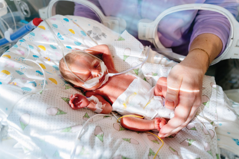
Extremely preterm newborns who weigh less than 3.3 pounds have immature lungs that often require high levels of ventilation oxygen in the hospital. This contributes to the chronic lung disease bronchopulmonary dysplasia, or BPD, the most common cause of death for these tiny infants. BPD exacts a devastating toll on the immature lung.

In one of the most extensive studies of the microorganisms in the intestines of very preterm infants, University of Alabama at Birmingham (UAB) and University of Tennessee Health Science Center researchers show that the gut composition of fungi in the second week of life predicts the later development of BPD, weeks to months before diagnosis of that disease. They analyzed gut fungi in the first true non-meconium stool produced before two weeks of life and found that the fungal intestinal microbiome—known as the mycobiome—of infants who later developed BPD differed in community diversity, composition and interconnectivity from the infants who never got BPD, as measured by the most up-to-date bioinformatic techniques. The researchers did not find significant differences in the bacterial microbiome in those first true stools.

To show causality, researchers transferred samples of the first true stool that predicts BPD or the first true stool of newborns who did not get BPD into female mice to give them a pseudo-humanized gut microflora. In a mouse model of BPD, newborn pups from those BPD dams showed an increased in the severity of lung injury compared with newborn pups from the no-BPD dams. In loss-of-function experiments, when the female mice with the BPD-predictive stool transplant were treated with an antifungal agent before birth, that inhibition of perinatal fungal colonization reduced lung injury in the newborn pups. In contrast, a gain-of-function experiment, where the perinatal fungal colonization of dams was augmented with a species of Candida fungus common in mice, amplified BPD severity in the newborn pups.

Kent Willis, M.D.

“These findings demonstrate that features of the initial intestinal fungal microbiome are associated with the later development of BPD in premature neonates and exert a microbiome-driven effect that is transferable and modifiable in mouse models,” said Children’s of Alabama neonatologist Kent Willis, M.D., who’s also an assistant professor in the UAB Department of Pediatrics Division of Neonatology. “This suggests causality, and it suggests that the gut fungi may represent a therapeutic target in newborn lung disease.”

Willis and Ajay J. Talati, M.D., University of Tennessee Health Science Center, Memphis, Tennessee, co-led the study, published in Microbiome.

“Collectively, our analyses demonstrate that the composition of the intestinal mycobiome of infants who did not develop BPD was more uniform,” Willis said. “In contrast, those who eventually developed BPD had more disparate mycobiomes. This suggests that a particular pattern of mycobiome development may be necessary to impart resistance to the development of BPD, and failure to do so in various ways is associated with disease development.”

The first defecations of newborn infants are meconium, composed from materials ingested while in the uterus. The first true stools in the second week, the ones analyzed by Willis and Talati, are digested milk. It is known, Willis and Talati say, that fungi in adults are vital members of the human microbiota; but compared to bacteria, their non-pathological and non-parasitic functions are still poorly understood, especially in newborns.

This prospective observational cohort study included newborn stool samples collected over six years from 2017-2020 in Memphis and 2021-2022 in Birmingham. The 64 very-preterm infants in the study who did not develop BPD had an average birthweight of 2.5 pounds, and the 38 very-preterm infants who did develop BPD had an average birthweight of 1.6 pounds. Only one of the 64 no-BPD infants in the study died, while six of the 38 BPD infants died.

Neonatology

A closer look at the effects of chorioamnionitis on premature babies

The majority of preterm births stem from chorioamnionitis. (Stock photo)

The vast majority of preterm births—especially “micro-preemies” born at 22 or 23 weeks’ gestation—stem from a single cause: chorioamnionitis, an inflammation of the placenta and membranes surrounding the fetus. But Children’s of Alabama neonatologist Viral Jain, M.D., is on a mission to determine why this insidious condition occurs, the ways it affects babies’ health, and how to stop it.

Occurring in an estimated 1% to 5% of births in the United States, chorioamnionitis—often shortened to chorio—can be hard to spot. It’s typically diagnosed using clinical signs of inflammation such as fever or elevated heart rates in either the mother or the baby. But chorio often eludes clinical diagnosis, silently causing damage to the placenta and triggering preterm birth, says Jain, also an assistant professor in the Division of Neonatology at the University of Alabama at Birmingham (UAB).

Viral Jain, M.D.

“It’s a huge reason why neonatology exists, as such,” he explained. “It’s the body’s reaction when there’s inflammation to deliver the baby preterm, and all the complications that come with a preterm baby are due to chorio. In addition, the inflammation also causes direct damage to the developing organs of the baby.”

Some of the extensive research conducted on chorio has focused on its causes, which may include infection, environmental chemicals, smoking and bleeding. But scientists still have a poor understanding of why it happens, Jain notes, as well as how to catch it early enough to stop premature delivery.

Much of Jain’s research has delved into chorio’s potential health implications for babies once they’re born—and the effects can be devastating. One of his studies shows that the incidence of cerebral palsy is far higher in infants born when chorio progresses to such a severe extent it becomes funisitis, or inflammation of the umbilical cord. Jain’s findings have been somewhat controversial, he acknowledges, since cerebral palsy is already known to affect more preterm infants than those born after full-term pregnancies.

“We chose the most severe chorio babies for the study to clearly show that it affects cerebral palsy development,” Jain said. “We found that it’s about 50-50—so half the risk of cerebral palsy was from being born pre-term due to chorio, and half was the direct injury coming from inflammation to the developing brain.”

To help predict the cerebral palsy risk of these infants while they’re still in the neonatal intensive care unit (NICU)—when early intervention can more easily be planned—Jain’s research has also used MRI to look for specific markers in the brain suggesting a high risk of the disabling condition.

“We showed that chorioamnionitis insult, which started at birth, continues in these babies and that we can see those changes in the MRI and that they lead to cerebral palsy,” he said. “This means you can start early intervention on those babies to capture or reduce some of the damage.”

Another of Jain’s studies suggests that infants born early due to chorio have chronic lung damage. “It creates an immune cell dysfunction in the lung that there is continuous damage happening,” he explained. In addition to requiring longer ventilator and oxygen treatment, these babies “end up developing what we call BPD, or bronchopulmonary dysplasia, which is neonatal chronic lung disease.”

Ultimately, Jain says, his research—which has been funded by the American Heart Association and National Institutes of Health—seeks to learn how chorio propagates so doctors can impede its damage.

“The goal is to find out what treatment we can give so when it’s just mild we can stop the progression and it won’t become full-blown chorio and end up delivering the baby preterm,” he said. “If we can do that, we can prevent a lot of organ damage to the lung or brain.”  

For more information on Jain’s work on chorio, listen to this episode of the Children’s of Alabama PedsCast podcast.

Neonatology

Using mitochondrial genetics to predict BPD

Researchers at Children’s and UAB are exploring how mitochondrial function may help predict BPD risk.

Bronchopulmonary dysplasia (BPD), a chronic lung condition affecting some extremely preterm infants, continues to be a significant clinical challenge in neonatology. While often lifesaving, supplemental oxygen can be a key contributor to long-term pulmonary complications in this vulnerable population. At Children’s of Alabama and the University of Alabama at Birmingham (UAB), researchers are exploring how mitochondrial function may hold the key to understanding and preventing BPD.

Jegen Kandasamy, M.D., an associate professor in the Division of Neonatology at UAB, leads a multidisciplinary team supported by a research grant dedicated to studying mitochondrial dysfunction in BPD. The research centers on individual differences in how mitochondrial DNA (mtDNA) haplogroups—genetic variations inherited maternally and varying by ethnicity—may influence an infant’s susceptibility to lung injury from oxygen exposure, particularly hyperoxia.

“Hyperoxia is a double-edged sword,” Kandasamy said. “It’s essential for survival, yet it introduces oxidative stress that preterm lungs are poorly equipped to handle. Our research is aimed at understanding how mitochondrial genetics impact that response.”

Using collected blood samples and clinical data from preterm infants, Kandasamy’s team is working to identify mtDNA haplogroups associated with higher BPD risk. The goal is to develop precise, genetically informed risk profiles that allow for early intervention. Hopefully, this will improve outcomes while addressing racial disparities in BPD prevalence and severity.

An especially promising area of research is platelet bioenergetics. By measuring how platelets utilize mitochondrial energy, the researchers hope to identify specific biomarkers that reflect systemic mitochondrial health and may help predict BPD risk. “Platelets are easy to access and give us a real-time snapshot of mitochondrial function without invasive procedures,” Kandasamy noted.

The team is also studying mitophagy, the elimination of damaged mitochondria through autophagy, and its role in lung development. Emerging evidence suggests that impaired mitophagy contributes to persistent mitochondrial dysfunction, exacerbating lung injury in preterm infants. As a result of this new evidence, the group is also evaluating the potential of thyroid hormone supplementation as a therapeutic strategy to restore mitochondrial function and mitigate lung damage.

By integrating clinical data with mouse models, the UAB team is uniquely positioned to investigate both the mechanistic underpinnings of BPD and potential interventions. The collaborative effort spans neonatology, mitochondrial biology and pediatric pulmonology, creating a comprehensive research environment.

“Our ultimate aim is to shift the paradigm from reactive to predictive personalized neonatal care,” Kandasamy said. “Understanding how mitochondrial genetics intersect with environmental exposures can help us identify at-risk infants earlier and intervene more effectively.”

Neonatology, Pulmonology

Improving lung function for COPD and BPD patients

A study led by Children’s researchers shows that inhalation of live Lactobacilli reduces inflammatory markers in BPD and COPD.

By Jeff Hansen, UAB

In preclinical models, the inhalation of a mixture of living Lactobacilli bacteria attenuated pulmonary inflammation and improved lung function and structure for the chronic lung diseases bronchopulmonary dysplasia (BPD) and chronic obstructive pulmonary disease (COPD).

This study, published in the journal Nature Communications, determined the mechanism of this live biotherapeutic product—a powder mixture of living Lactobacilli bacteria—to reduce neutrophilic inflammation and reduce a broad swath of inflammatory markers in BPD and COPD, says Charitharth Vivek Lal, M.D., a neonatologist at Children’s of Alabama and the University of Alabama at Birmingham (UAB). Lal co-led the research with Amit Gaggar, M.D., Ph.D., a UAB pulmonologist.

Their findings “provide a paradigm for the progression of structural lung disease,” Lal said, because they identify the Lactobacilli as critical to regulating lung protease activity that is linked to the destruction caused by matrikine generation, extracellular matrix turnover and chronic neutrophilic inflammation that damages air sacs in the lungs. 

A possible protective role for Lactobacilli in the lung and the possible use of Lactobacilli to treat chronic lung disease had its foundation in 2016 when Lal and UAB colleagues discovered that the airways of infants with severe bronchopulmonary dysplasia had decreased numbers of Lactobacilli, increased numbers of proteobacteria and increased concentrations of proteobacterial endotoxin. In this latest study, the UAB researchers provided a mechanism of action for the Lactobacilli treatment to decrease downstream disease development and showed safety and effectiveness of the live biotherapeutic treatment in a mouse pup model for BPD and three mouse models of COPD. 

Bronchopulmonary dysplasia develops in some extremely premature infants after damage induced by high oxygen tension or mechanical ventilation needed to keep them alive. COPD occurs in older people, especially smokers, and kills about 130,000 Americans a year and about 3 million more worldwide.

“Inhaled live biotherapeutic products show promise in addressing common pathways of disease progression that in the future can be targeted at a variety of lung diseases,” Lal said. “Preclinical animal data is suggestive, and safety of the potential drug in humans will be tested in a forthcoming clinical trial. Human adult safety data in COPD will help de-risk the pathway to approval for use of the drug in bronchopulmonary disease infants.”

The UAB researchers hypothesized that mouse models of BPD would show heightened levels of acetylated proline-glycine-proline, or Ac-PGP, an extracellular matrix-derived peptide, as had been seen in premature infants with BPD.

This was demonstrated in BPD mouse models, and gain- or loss-of-function studies showed the impact of Ac-PGP. Intranasal instillation of Ac-PGP increased neutrophilic inflammation and lung degradation. When an inhibitor of Ac-PGP was given with the Ac-PGP, markers of neutrophilic inflammation decreased and lung structure improved.

Researchers then showed that a proprietary Lactobacilli blend of L. planatarum, L. acidophilus and L. rhamnosus performed best in synergy to reduce the inflammatory proteinase MMP-9, which helps release the Ac-PGP from extracellular matrix. Furthermore, supernatant from Lactobacilli growth medium also reduced MMP-9 at a similar magnitude as live Lactobacilli bacteria. 

A key finding was that L(+) lactic acid, which is produced in Lactobacilli growth medium supernatant, reduced MMP-9 in vitro, showing an important role for this lactic acid as an anti-inflammatory molecule. Researchers found that live Lactobacilli in the lungs provided an ongoing, sustained release of L(+) lactic acid in a controlled and well-tolerated manner.

A major technological advance reported in the study was creating the inhaled Lactobacilli powder through particle engineering—particles small enough to reach deep into the lungs while preserving viable bacteria. This live biotherapeutic product was then tested in the BPD and COPD models. In the COPD mouse models, the blend successfully reduced inflammation in the lung microenvironment whether treated concurrently or post-injury, showing anti-inflammatory effects, decrease of several pro-inflammatory markers and elevation of the anti-inflammatory marker IgA. 

An interesting finding was the favorable performance of the live biotherapeutic product. It reduced MMP-9 and other pro-inflammatory cytokines as effectively as—or in some cases better than—fluticasone furoate, a United States Food and Drug Administration-approved inhaled corticosteroid found in COPD combination therapies. 

Safety and biodistribution studies in one of the COPD mouse models showed that inhalation of the bacterial powder did not initiate adverse reactions or disease, and the Lactobacilli did not translocate to distal tissues or accumulate in the lungs.

Neonatology

Uncovering the Role of the Pulmonary Microbiome in Chronic Respiratory Disease

Say the word microbiome and you probably think about the billions of microbes that inhabit the gut. But Children’s of Alabama neonatologist Charitharth Vivek Lal, M.D., wants you to consider another microbiome — the lung microbiome. Not only does it exist, he and his team have discovered, but it is present as early as birth, even at 24 weeks gestation, negating the long-held believe that the lungs are sterile before birth.

The question he is now trying to answer is what role it plays in the chronic lung disease bronchopulmonary dysplasia (BPD), which affects between 48% to 68% of babies born before 28 weeks of gestation. The condition is a major cause of morbidity and mortality in preterm infants, characterized by lung inflammation, injury and pulmonary hypertension, among other factors.[1]

A study from Lal clearly demonstrated that microbial imbalance, or dysbiosis, predicts the development of BPD in extremely low-birthweight newborns. He and his team evaluated the microbiome of several infants at birth and found diverse and similar airway microbiomes in both, which differed from older preterm infants with BPD.

They found that dysbiotic changes in the airway microbiome at birth correlated with the development of BPD, including lower levels of the “good” bacteria lactobacillus in infants born to mothers with chorioamnionitis, an infection of the membranes of the placenta that is an independent risk factor for BPD. They suggested in their paper that a microbiome signature possibly exists in utero, and that part of its role may be to prime the pulmonary immune system. If dysbiosis occurs, they wrote, “it may set the stage for subsequent lung disease.”

So, said Lal, what about a respiratory probiotic to restore the microbiome?

“If it relieves inflammation, could we use this to replace steroids in various childhood lung diseases?” he asked. Studies in mice using Dr. Lal’s patented ‘respiratory probiotics’ demonstrate benefits. “The next step is to test it in larger animals and then humans,” he said.


[1] Lal CV, Bhandari V, Ambalavanan N.Genomics, Microbiomics, Proteomics and Metabolomics in Bronchopulmonary Dysplasia. Semin Perinatol. 2018 Nov;42(7):425-431.

[2] Lal CV, Kandasamy J, Ramani M, Ambalavanan N. Metabolomic and Metagenomic Signatures of Bronchopulmonary Dysplasia. Am J Physiol Lung Cell Mol Physiol. 2018 Aug 16.

[3] Lal CV, Olave N, Travers C, Halloran H, Rezonzew G, Xu X, Genschmer K, Russell D, Gaggar A, Blalock E, Vineet Bhandari, Ambalavanan N. Exosomal MicroRNA 876-3p Predicts and Protects Against Severe Bronchopulmonary Dysplasia in Extremely Preterm Infants. JCI Insight, 2018; 3(5: e93994). PMID: 29515035

Care for the Tiniest Patients

Learn more about the Department of Neonatology at Children’s of Alabama.