Browsing Tag

cardiology

Cardiology, Inside Pediatrics

Lau shares vision as new Physician-in-Chief

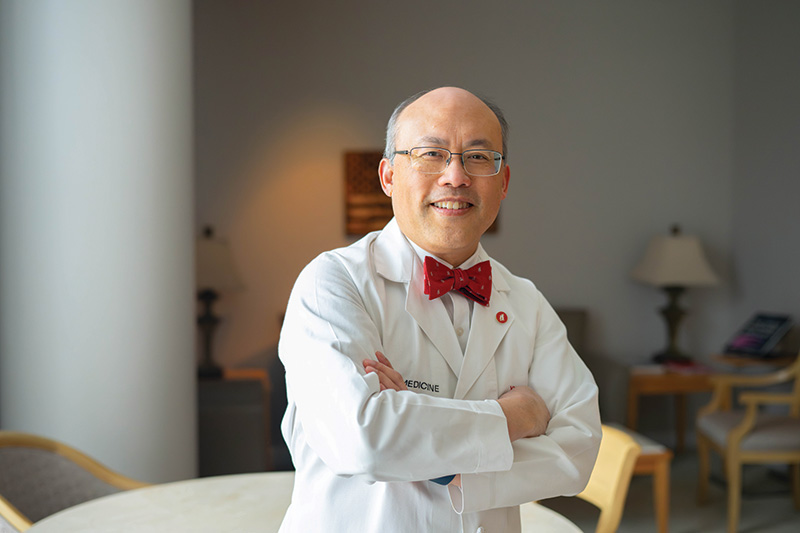
Yung Lau, M.D. was named physician-in-chief at Children’s of Alabama and chair of the UAB Dept. of Pediatrics in March 2025.

After serving as interim chair for five months, Yung Lau, M.D., was officially named chair of the University of Alabama at Birmingham (UAB) Department of Pediatrics and physician-in-chief at Children’s of Alabama in March 2025. The only real change, Lau said, was that he could now formally begin planning for the future. His vision for the department is expansive—centered on collaboration and faculty support. But he believes the path to those big goals lies in the small things everyone does every day.

Lau stepped into the interim chair role in November 2024, following the announcement that then-chair Mitch Cohen, M.D., would be departing at year’s end to join Stanford. “Dr. Cohen led for a decade and helped build this department into a strong and vibrant group,” Lau said. “It has consistently thrived, and I’ve considered it a privilege to be a part of the department as a faculty member for over three decades. Now, it’s an awesome responsibility to carry on this tradition of excellence.”

The role requires close collaboration between two major institutions: Children’s of Alabama and the University of Alabama at Birmingham. With more than 30 years of experience across both organizations—including in leadership roles—Lau understands their individual missions and how they intersect.

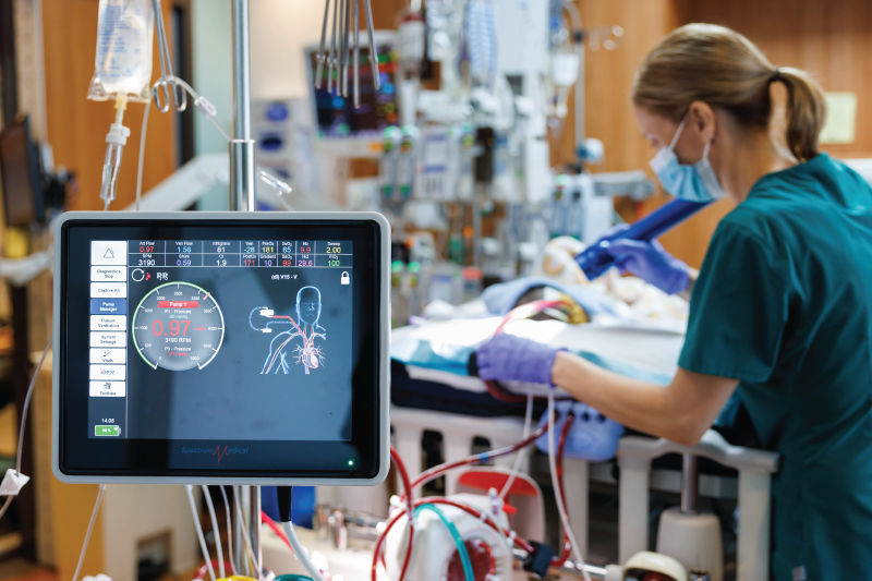
That understanding came into sharp focus in 2007, when Lau led the UAB group in a major collaborative effort between the two institutions. At the time, UAB housed the pediatric cardiac program. But as Children’s leaders planned to build a new hospital tower, they wanted to bring the program under their roof. Over the next five years, Lau played a significant role in bringing physicians and other clinical staff to assist in the design of the cardiac intensive care units, operating rooms, catheterization labs, step-down units and cardiovascular perioperative areas. He then worked closely with Children’s leadership on staffing and operations planning. On October 14, 2012, the program moved into the new building—seamlessly.

The success of the move laid the foundation for a quantum leap in the ability of the Pediatric and Congenital Heart Center of Alabama to provide state-of-the-art care. Today, the program consistently ranks among the top performing pediatric and congenital heart centers in the nation by numerous metrics, and the Society of Thoracic Surgery has classified the program as an overperforming center—one of 12 in the country. For Lau, who served as the division director of pediatric cardiology from 2012 until his appointment as chair in 2025, the experience left a lasting impression about what’s possible when Children’s and UAB work together.

“The opening of the Heart Center marked one of the most satisfying periods of my career,” he said. As he steps into his new role, he hopes to lead more collaborative efforts with similarly meaningful impact.

The Heart Center’s success is a powerful example of the synergy between Children’s and UAB—a synergy Lau believes can grow even stronger. “What I’ve seen is a real willingness among leadership across both institutions to reduce barriers and connect silos,” he said.

Lau has outlined three key priorities to strengthen that collaboration: maximizing current resources in clinical care, education and research; strategically recruiting and developing faculty; and building a resilient foundation of financial stability and physician well-being.

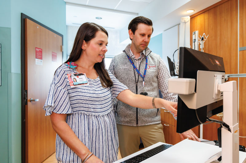
Education and research are crucial parts of his strategy, and UAB and Children’s have a history of successful collaboration on both. From an education perspective, Children’s serves as the teaching hospital for the UAB pediatric medicine, surgery, psychiatry, research and residency programs. “There’s this deep core of understanding between Children’s and the department that we are really training the future physicians for the state,” Lau said. And that’s a crucial role in a state that, Lau says, needs more physicians and pediatricians. “Part of our duty here is in our obligation to do that,” he added.

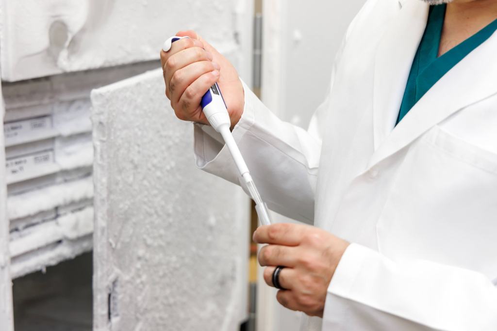
On the research side, the two institutions work together to “advance knowledge for the children of Alabama and beyond for the future,” Lau said. This benefits both entities, sometimes leads to advancements and breakthroughs that influence the broader world of medicine, and enhances the reputation of both.

“When we collaborate more extensively and continue to strengthen those ties of collaboration, two important things happen,” he said. “First, children receive better care, now and in the future. Second, our faculty experience significantly greater job satisfaction.”

Faculty support is another central pillar of Lau’s vision. Since becoming chair, he has met with many faculty members—some he’s long known, others he’s come to know better through these conversations. What stands out most, he says, is their passion and the profound impact they have on children’s lives. His goal is to listen, support and help them succeed.

Lau also acknowledges the tension between moral obligation and financial reality. “That’s just medicine in America today,” he said. But he’s confident the department can thrive within that framework.

“I think both institutions understand that we need to maximize our resources—our people and infrastructure—to provide the best possible care, to train the next generation of pediatricians, and to innovate through research,” he said.

Though there are multiple facets to Lau’s vision, everything is focused around the patient.

“The patients in front of us are the cornerstone of everything we do,” he said. “And while our goals may be big, the real progress happens in the small steps we take every day.”

“Yes, having a goal is important,” he continued. “But sometimes if we focus only on the goal, we risk losing sight of what’s happening in the moment—and that can distort the work being done on the ground. Sometimes the things that matter most get sidelined in the name of progress.”

With a strong focus on the patient—and through strong collaboration and faculty support—Lau believes UAB and Children’s will continue to deliver exceptional care to every child they serve.

Cardiology

Law appointed division director of pediatric cardiology

Mark Law, M.D., is the new director of the Division of Pediatric Cardiology at Children’s of Alabama and UAB.

By Heather Watts (UAB)

The University of Alabama at Birmingham (UAB) Department of Pediatrics announces with great pleasure and gratitude the appointment of Mark Law, M.D., professor in the Department of Pediatrics, to the permanent position of director of the Division of Pediatric Cardiology at UAB and Children’s of Alabama. Since stepping into the interim role in November 2024, Law has made substantive changes that address current needs as well as laying a strong foundation for continued growth and success within the division and the Pediatric and Congenital Heart Center of Alabama at Children’s of Alabama. Law brings his remarkable thoughtfulness paired with a blend of clinical excellence, research innovation and mentorship to this leadership role.

“In a twist of irony, when I interviewed Dr. Law to join us in 2008, he confidently declared that he had no interest in becoming a division director,” said Yung Lau, M.D., professor and chair of the Department of Pediatrics and Law’s predecessor as division director. “Fortunately, his thinking evolved. When the intersection of his unique talents and the Heart Center’s greatest needs became clear, he answered the call.”

Over the years, Law has progressed through the academic ranks—from assistant professor to his current role as professor of pediatrics. He also serves as medical director of Adult Congenital Interventional Cardiology at UAB Medicine, with a secondary appointment in the Division of Cardiovascular Disease within the Department of Medicine.

Widely respected as a leader in pediatric and interventional cardiology, as well as adult congenital heart disease, Law has authored or co-authored more than 70 peer-reviewed articles and book chapters. He has also mentored more than 20 post-doctoral fellows and junior faculty, contributing meaningfully to the future of academic medicine.

In recognition of his many accomplishments and unwavering dedication, Law was recently appointed to the prestigious Lionel M. Bargeron Endowed Chair in Pediatric Cardiology by the Board of Trustees of the University of Alabama—a most fitting appointment given the pioneering contributions Bargeron made to the field of heart catheterization when it was in its infancy.

Cardiology

Children’s of Alabama’s interstage home monitoring program growing

Brittany Abercrombie, NP, and Alan Brock, M.D., discuss the progress of a patient in the Hearts at Home program.

As the reputation of the Children’s of Alabama Pediatric and Congenital Heart Center of Alabama has grown, so has the success of its programs. Case in point—Hearts at Home, an interstage home monitoring program for any patient with single ventricle physiology who has undergone their first palliation procedure. In the last five years, the program has seen steady growth in the number of these patients, and leaders say the center’s reputation is among the reasons why.

“I think as a heart center in general, we’ve just had an influx of patients,” said Brittney Abercrombie, a nurse practitioner and the coordinator of Hearts at Home. “And so by default, that means that we are having more interstage patients.”

When Abercrombie moved into her role five years ago, Hearts at Home was caring for six to eight patients at a time. Now, she says they typically have about 13. Yearly, the program follows as many as 30, compared with 23-25 when she began. In the last couple of years, they’ve attracted more patients from outside Alabama, including children from Georgia, Tennessee and the Pensacola, Florida, area. Some of the program’s patients chose Children’s over other options in the region.

“I think they recognize that our outcomes here are some of the best in the Southeast,” said Alan Brock, M.D., the program’s medical coordinator. “And when they have the opportunity to look around and pick which program they want, I think patients are choosing us.”

As a result of the program’s success, hypoplastic left heart syndrome—a condition that brings many patients to the program—has become one of the most common forms of single ventricle congenital heart disease the hospital treats, Brock added. “I think it’s because we’re getting better at what we do and we’re saving a lot more lives now,” he said. “That is part of the reason that there are more patients coming into our program.”

What is Hearts at Home?

Through the Hearts at Home program, the families of patients with hypoplastic left heart syndrome and other forms of single ventricle congenital heart disease have access to education and technology that helps them to monitor and track their child’s heart health at home during the period between their first and second stages of palliation—procedures designed to repair their congenital heart defect. This time is tenuous for the child and often stressful for the parents, requiring a great deal of medical management, including monitoring, medications, adhering to strict feeding regimens, checking vital signs and having emergency access to equipment. “I think especially for these first-time parents, they don’t know what’s normal and what’s not,” Abercrombie said. “They’re not only learning to parent, but they’re learning how to parent a medically fragile child, so I think that’s a big challenge for them.”

There’s also the threat of morbidity, which is what led to the creation of interstage monitoring programs. The effort began in 2008 with the formation of the National Pediatric Cardiology Quality Improvement Collaborative (NPC-QIC). Since then, interstage monitoring programs across the country have succeeded tremendously, dropping the interstage mortality rate by more than 40%, Brock said.

The programs are effective because of their focus on education, data and communication. The work begins before a family even leaves the hospital. While there, they go through extensive training to help them understand their child’s condition, how to manage it and the warning signs that might arise. Once they’re home, they track all of their child’s vitals—specifically heart rate, oxygen and saturations—through an app called Locus Health. This data is accessible by the patient’s care team, giving them a look at the patient’s trends and helping them to quickly identify any problems. “It helps us see the whole picture while they’re at home,” Abercrombie said. If any issues do arise, the family can connect with the care team via messages through the app, and providers can even use the app for telehealth appointments, if necessary.

In one case at Children’s, monitoring may have saved a child’s life. Abercrombie says the team detected a change in heart rate and some feeding intolerance, which, combined with the patient’s trends, indicated they needed medical attention. The team called the mom, got the patient in for a visit and prevented a medication overdose. “If we didn’t have [the monitoring], there’s a good chance that could have ended up in a mortality,” Abercrombie said.

The team

The Hearts at Home team includes, in addition to Abercrombie and Brock, cardiologists who see most of the interstage patients, a nutritionist who specializes in cardiovascular disease, a social worker and speech therapists. Nurse practitioners or intensivists are available to answer parents’ questions 24 hours a day, which can be reassuring. “It is just a very small group of people that are caring for these patients day in and day out, along with the family,” Abercrombie said. “And so I think that they feel a lot of comfort and confidence in knowing that there’s someone there to talk with them and help them throughout the day.”

This frequent communication can lead to close relationships between the parents and the care team—so much that when the child eventually “graduates” from the program (after having their second palliation procedure) and no longer has the same level of access to the team, the achievement is often bittersweet.

“It’s a good thing,” Abercrombie explains to the parents. “It means your baby has a much more stable heart. You shouldn’t need us as much. They can do a lot more normal baby things.”

“But [the parents] do have a little bit of sadness about losing kind of that access,” she added.

When a new patient enters the program, the team contacts their pediatrician to share information about the patient’s condition and explain how the program works and what to expect. They also reach to local EMS in the patient’s community to inform them that a congenital heart disease patient lives nearby so they’ll be prepared in case there’s ever an emergency.

Going forward, the program may expand to older patients. Brock hopes to focus future efforts on neurodevelopmental outcomes and “how these kids develop throughout the course of their single ventricle life,” he said. Nationally, the NPC-QIC recently merged with the Fontan Outcomes Network to form Single Ventricle One (SV-ONE) in an effort to follow these patients beyond their palliation procedures into their teens and beyond.

Cardiology

Flow restrictors offer hope for high-risk newborns

Flow restrictors, like the one pictured, are showing promise for helping patients with hypoplastic left heart syndrome.

Babies born with hypoplastic left heart syndrome, in which the left side of the heart doesn’t fully develop and thus can’t pump blood very well, typically require open-heart surgery soon after birth or a hybrid surgical approach combining stenting and open surgery to establish systemic blood flow and control pulmonary blood flow. Given the already fragile state of the infant, however, neither approach is optimal, and both have high mortality rates.

Doctors at Children’s of Alabama are helping these children by using an internal pulmonary flow restrictor created by modifying a microvascular plug. They form the device by cutting a tiny hole in the Gore-Tex covering of the microvascular plug originally designed to close blood vessels in children and adults. With this use of this device, they are able to postpone surgery for months until the infant is bigger and stronger while still controlling pulmonary blood flow in a completely non-invasive manner.

“The problem we’re addressing is over-circulation of blood to the lungs,” explained pediatric cardiologist Mark Law, M.D. “Traditionally, it requires open-heart surgery to place a band around the pulmonary artery to reduce blood flow. However, this surgery is invasive and can be very stressful for the baby.”

Now, Law and his team are able to treat the infant in the cath lab rather than an operating room. There, they thread the plug through the femoral or internal jugular vein into the pulmonary artery to partially restrict excessive blood flow to the lungs. “It’s a far less invasive procedure and the recovery is faster,” he said. It also allows the surgeons to operate without scar tissue from a previous surgical procedure.

This procedure enables babies who are too fragile for surgery to safely grow stronger before undergoing their first operation. In one study, 10 out of 13 infants treated with this technique survived to their next surgery, typically performed within two to six months of the catheter procedure. When compared to historical data from similar high-risk patients who underwent traditional procedures, the new technique was associated with significantly better six-month survival rates after adjusting for other risk factors.[1]

Importantly, the baby’s pulmonary arteries grow over that time, which is crucial for their long-term outcomes. In addition, the devices can be easily removed during subsequent surgical procedures.

Law and his team have presented a series of five cases in which they used the technique on babies who were too medically fragile for surgery. All survived and all were later able to undergo the needed open surgery. “We’re confident some of these babies wouldn’t have made it otherwise,” he said. “But because they’re older, bigger, stronger, and breathing on their own,” the procedure was much less risky.

Still, challenges remain. The device must be customized for each patient, and its small size limits its use to very young infants.

Nonetheless, pediatric cardiologists across the country are adopting the procedure, Law said. “We’re all learning from each other and sharing data to refine the technique.”

“This technique represents a shift in how we think about treating congenital heart defects,” Law said. “The ability to delay surgery and avoid scar tissue is a game changer.”


[1] Sperotto F, et al. Circ Cardiovasc Interv. 2023;16:e013383. DOI: 10.1161/CIRCINTERVENTIONS.123.013383

Cardiology

Exercise Testing for Fontan Patients

Cardiologists at Children’s of Alabama are using exercise training to measure Fontan patients’ health.

Children born with single-ventricle heart disease undergo multiple surgeries before age 6, ending with the Fontan procedure, which redirects blood from the inferior and superior vena cava to the pulmonary arteries. This allows blood returning from the body to skip the overworked single ventricle and instead flow directly to the lungs to receive oxygen. While the surgeries have improved survival for these patients, long-term outcomes are still poor. Now, clinicians at Children’s of Alabama are honing in on exercise as an important way to track and improve their quality of life.

“The Fontan operation is one of the most complicated congenital heart procedures we do,” pediatric cardiologist Camden Hebson, M.D., said. “Even with the best outcomes, there’s a shortened lifespan. Many patients start to have major complications by the time they’re in their 20s and 30s, such that it’s pretty unusual for patients to be still living, especially symptom free, in their 50s.”

Camden Hebson, M.D.

The fundamental challenge lies in the procedure’s physiology. In normal hearts, the right side actively pumps blood to the lungs. Because the Fontan operation eliminates this pump, it creates a passive flow system that, while life-sustaining, leads to increased venous pressure throughout the body, particularly in the abdomen and legs.

“The pie in the sky for these patients is getting some sort of pump into them to better push the blood to the lungs,” Hebson said. “There’s all sorts of ideas out there of how to do that, like mechanical implantations and valves. But human beings have our own intrinsic pump that we can maximize: the skeletal muscle pump in our legs, which push venous blood out of the legs back up toward the lungs.”

People who don’t exercise a lot have weak skeletal muscle pumps, he said, which increases the blood pooling and pressure on vital organs, resulting in complications and early mortality. “People who have the strongest legs, the most physically fit legs, the best muscle tone are going to actuate their skeletal muscle pump as much as possible,” he said.

Yet, families and medical teams have traditionally taken a protective approach with these children and limited physical activity, Hebson said. “In some ways, that’s actually a negative, because then these kids are less active than the other kids their age,” he explained. “They exercise less, and then it actually makes the problems of the physiology more likely to occur in their 20s and 30s.”

The link between exercise and better outcomes comes from studies on long-lived Fontan patients called “Super-Fontans.” While the exact reasons for their success aren’t fully understood, physical fitness appears to play a crucial role.

To better understand the effects of exercise on the cardiovascular system of Fontan patients, Hebson and his team use metabolic exercise testing to track heart rate and blood flow during physical activity. This not only helps identify potential treatment targets and provide reassurance about exercise safety, but serves as an early warning sign for complications. For instance, it can reveal issues that aren’t usually apparent at rest, such as the need for stenting the Fontan conduit based on limitations shown in exercise capacity.

“Sometimes, you just need to take the car out on the road and see how it drives,” he said, comparing traditional resting assessments to testing patients during exercise. “For most patients, in particular Fontan patients, your cardiovascular system is what limits your ability to exercise. So how much exercise you can do at any given time is really just feedback on how well the heart works.”

“So we need to encourage our patients to be ahead of the curve on how much exercise they do to keep their legs strong,” Hebson continued. “Not only are they getting the benefits that everybody gets from exercise, they are actually improving the venous return to their heart and their cardiac output as a result.”

“Anything that can be done to improve these patient’s lives and long-term outcomes would be a big deal,” he added.

Cardiology

Saving lives in Latin America

Each year, a team from Children’s of Alabama travels to Peru to provide cardiovascular surgeries through Heart Care International.

For Children’s of Alabama cardiac pediatric intensivist Santiago Borasino, M.D., traveling to Peru each year as part of a team providing cardiovascular surgeries to children offers him a special way to give back to his home country.

The trips are sponsored by Heart Care International, a nonprofit organization committed to saving the lives of children born with congenital heart defects. Volunteer medical teams come from cardiac centers throughout the United States, including Children’s of Alabama, to provide care throughout Latin America.

Borasino’s first trips were to Chiapas, Mexico, but since he learned about the organization’s work in Peru, he has focused his efforts there.

Santiago Borasino, M.D.

“I get to give back to my country,” he said. “There’s always a lot of guilt for leaving. And so I get to go there, help with surgeries and give back to the country some—a little bit. This teeny tiny bit probably doesn’t repay everything that the country has done for me, but it’s a little bit. It’s very special to me.”

The team—which most recently included Children’s cardiothoracic surgeon Robert Sorabella, M.D.; pediatric cardiac anesthesiologists Jack Crawford, M.D., Ph.D., and Patrick Hussey, M.D.;  and Stephanie McBride, RNFA—works with doctors at the Instituto Nacional de Salud del Niño (INSN) San Borja in Lima, where Borasino was raised and attended medical school.

The local doctors coordinate with the visiting clinicians to choose the cases. “We can’t do cases that are too complex because we don’t have ECMO,” Borasino said. ECMO, or extracorporeal membrane oxygenation, is a life-support machine that can temporarily replace the heart and lungs. They also want the child to be able to recover before the team leaves, so they have to choose wisely, he added.

The visiting team works closely with their Peruvian counterparts. The American and Peruvian surgeons, anesthesiologists and nurses team up for surgeries, while Borasino partners with his Peruvian counterpart to manage the post-operative care side in the ICU. They generally perform about 15 surgeries a day.

Over his six trips to Peru, Borasino has witnessed significant growth in the skills and knowledge of the local medical professionals, most of whom were just starting to practice when he met them. “They’ve grown just like any doctor in the United States grows from being a young doctor who hasn’t done this too much to somebody who’s done it quite a lot.”

“The ultimate goal is that they will be independent,” he said. “They’re never going to be like Americans because of the resources. But, in theory, they could get close, at least for the moderate or low cases, a little complex but not too complex.”

Without these volunteer trips, the outlook for many of the young heart patients is grim. “Some of them will be lucky enough to get surgery within the system,” Borasino said. “And some of them will die. Not immediately, but eventually. Like in a few years without surgery, you end up dying. All these congenital heart diseases eventually kill you, either in weeks, months or years.”

However, the team’s work can be life-changing for the patients they’re able to help.

“The families are so thankful,” Borasino added. “Some come back every year when they know we’re there just to say hi, to bring their kids and tell us, ‘You saved our kid’s life.'” “It’s an opportunity to see more than just cardiac medicine,” he added. “And just to help.”

Cardiology

Heart team discovers new lung injury biomarker

Ahmed Asfari, M.D., with a patient in the Pediatric & Congenital Heart Center of Alabama at Children’s of Alabama.

Nearly all children who undergo cardiac surgery suffer lung injury afterward, and its treatment can determine whether the injury is short-lived or will follow a child for a lifetime. That’s why the discovery at Children’s of Alabama of a new biomarker to predict which patients are at higher risk of this complication—steering the use of respiratory support—is being heralded as a groundbreaking development that may reap benefits for children far and wide.

Using samples from a Children’s biorepository, heart specialists revealed that blood levels of a protein called proteoglycan 4, commonly known as lubricin, significantly differ between children undergoing long- and short-term mechanical ventilation after cardiac surgery. Until now, physicians haven’t been able to predict which patients stood at higher risk of lung injury in this care setting.

It’s the first time the finding has been reported in the United States, said Ahmed Asfari, M.D., a cardiac intensivist at the Pediatric & Congenital Heart Center of Alabama at Children’s.

“There’s been nothing like this in regards to acute lung injury, especially for our patient population with congenital heart surgery,” said Asfari, who’s also an assistant professor in the Department of Pediatrics and Division of Cardiology at the University of Alabama at Birmingham (UAB). “When we tested the blood of patients with very long mechanical ventilation duration and compared them to patients with short duration, we found the level of this marker goes down, especially within two days of surgery.”

The discovery wouldn’t have been possible without the Children’s biorepository (left), which houses samples dating back over a decade. All patients under age 8 admitted for heart surgery had blood collected at several points in care, including before and after heart and lung bypass treatment, contributing to this research. The findings were validated at the Dr. Tannin Schmidt Lab at the University of Connecticut and published in Translational Pediatrics.

Asfari said the breakthrough likely will change the landscape for children’s care in the future. “Having the ability to have a serum biomarker that we can use to grade the level of acute lung injury will be extremely helpful,” he said.

The next stage of research will also expand insight into how widely the biomarker may be used. Blood testing now includes larger numbers of patients, along with those in different age groups and with varying cardiac physiology and anatomy, as well as those undergoing other types of surgery, and those both on and off bypass.

“The next step of our research will be doing it prospectively, looking at the patients, healthy children, and also patients with acute lung injury with different physiology—not just heart disease—and over a longer period,” Asfari said.

If the results hold, he predicts the new biomarker might one day become a gold-standard test to predict patients’ odds of suffering acute lung injury.

“It has very good potential to be used at bedside,” Asfari said. “This kind of study and research is very important to adjust the care we’ll provide for each individual patient and shines a light on the role of customized medicine for the future.”

Cardiology

The benefits of bivalirudin during ECMO care

A new study is shaking up the standard practice of using heparin for anticoagulation in ECMO patients.

Anticoagulation therapy during extracorporeal membrane oxygenation (ECMO) has long relied on heparin, but a groundbreaking multicenter study co-authored by Jonathan Byrnes, M.D., medical director of cardiac ECMO at Children’s of Alabama, shows the benefits of replacing heparin with bivalirudin.

Bivalirudin isn’t a new drug, but its rise in use in pediatric ECMO stems from successes in pediatric ventricular assist devices (VADs). With earlier stroke rates as high as 25–30% on VADs using heparin, the transition to bivalirudin brought about a remarkable reduction, dropping the stroke rate to 5–8%. This significant improvement in neurological outcomes sparked interest in applying bivalirudin more extensively in ECMO settings.

The study, spearheaded by Mohammed Hamzah, M.D., of Cleveland Clinic Children’s Hospital, diverged from past practices of individual center decision-making. Instead, six medical centers collaborated, aiming to analyze outcomes through a case-matched lens, minimizing variability across patients requiring ECMO. The study’s cooperative nature across multiple centers necessitated remarkable coordination.

One of the study’s key findings, according to Byrnes, is that patients on bivalirudin exhibited lower mortality rates than those on heparin. Intriguingly, despite higher PTT levels in bivalirudin patients, they experienced less bleeding and fewer thrombotic incidents in the ECMO circuit. This hints at bivalirudin’s inherent benefits, possibly enhancing biocompatibility. “It seems that there’s something intrinsic to bivalirudin that allowed for better biocompatibility of the patient’s blood to the ECMO circuit,” Byrnes said.

The impact of this study extends far beyond the academic realm. At Children’s of Alabama, the findings suggest potential benefits such as reduced bleeding, fewer transfusions and lower mortality rates for ECMO-supported patients. However, as Byrnes noted, the retrospective nature of the study necessitates a randomized controlled trial to solidify these outcomes and effectively influence the standard of care.

Plans for a prospective trial are underway, aiming for a randomized approach coordinated through the Extracorporeal Life Support Organization (ELSO). This prospective study could validate the retrospective findings, potentially reshaping protocols for anticoagulation during ECMO.

Despite the excitement surrounding the study’s outcomes, Byrnes emphasized the need for caution. While retrospective data suggests bivalirudin’s superiority, a randomized trial is crucial to establish this definitively. Prospective data will unveil any center-specific confounders that might have influenced the study’s retrospective findings.

Byrnes underscored the significance of these findings and the hope for future randomized controlled trials. The study’s retrospective nature lays a robust foundation, but prospective data will offer irrefutable evidence to guide clinical practice, leading to a potential paradigm shift in anticoagulation therapy that may result in improved patient care and outcomes.

Cardiology

Code Committee Brings Innovation and Improvements

HC3 is led by Dr. Ahmed Asfari (top row, fourth from left) and Ashley Moellinger (top row, third from left).

Since the inception of the Heart Center Code Committee (HC3) in 2014, Children’s of Alabama’s cardiac arrest rate in the Cardiovascular ICU has fallen nearly 50%. The impact this committee has had on cardiac arrest reduction has come from numerous quality improvement initiatives and safety changes. In 2022, some of the initiatives Children’s implemented include the development of four guidelines, three communication enhancement tools, checklists and numerous safety changes.

Whenever a patient goes into cardiac arrest or a near miss is encountered, HC3 discusses the case. This multidisciplinary committee is composed of physicians, nurse practitioners, bedside nurses, respiratory therapists, cardiovascular operating room staff, chaplains and leadership from the heart center. HC3 meets every other week to evaluate each case, identify what was done well and areas for improvement.

Nurse practitioner Ashley Moellinger, CRNP, MSHQS, and cardiac intensivist Ahmed Asfari, M.D., who participate in quality improvement initiatives within the heart center, began leading the committee in 2021. They’ve focused on three key areas: education, inclusivity and innovation.

“We have unique patients with really complicated conditions that can be challenging to understand,” Moellinger said. “Whenever we review the event, we look for areas where there’s a knowledge gap or a need to enhance skills.” Then the team sends education briefs to the nurses to highlight committee findings.

Every nurse in the department is involved with the committee. “Really, the committee is owned by the nurses,” Moellinger said. An elite team of nurses reviews each case, interviews those involved and develops a presentation to tell the story of the event. Then, the committee decides together what they need to change. “And that’s where the education rollout comes into play,” Moellinger said.

The committee uses artificial intelligence and near-real-time analytic algorithms to analyze cardiac arrest and near-arrest events. “When you’re talking about a cardiac arrest, everyone’s recollection is going to be different,” Asfari said. With the platform, “we can use objective data to show the patient’s course.” It also allows the team to view vital signs in a continuous manner on one screen.

“One of the things we are most proud of is the ‘green’ epinephrine action plan,” Asfari said. When a patient goes into cardiac arrest, epinephrine is one of the first medications administered, but it takes time to prepare the drug. Shorter time to administer it is associated with better outcomes. The action plan calls for prepared epinephrine at the bedside and includes standing orders for the nurse to administer it once the patient’s vitals reach a certain threshold. “It’s made a huge impact on our patient care,” he said. “Cardiac arrest is a real problem for children with cardiac disease because they are so fragile,” Asfari said. “Improving resuscitation and, more importantly, preventing the arrest can improve the outcome.”

Cardiology

New Registry Studying Long-Term Outcomes of Heart Procedures

A new national registry is tracking patients’ progress over time following heart procedures as neonates.

Children’s of Alabama has joined a new national registry managed by the Congenital Cardiac Research Collaborative (CCRC), attempting to answer the question: What is the best way to treat ductal-dependent pulmonary blood flow congenital heart conditions?

Conditions such as Tetralogy of Fallot, critical pulmonary valve stenosis and complex single-ventricle arrangements can be treated with various interventions, both percutaneous and surgical. “We’re looking at all neonates (first 30 days of life) who require a procedure, so they get enough pulmonary blood flow to survive,” Children’s pediatric cardiologist Mark Law, M.D., said. “The goal is to take this broad grouping of patients and understand how they do, not just with their first intervention or even their second intervention if they need one, but how do they do as they grow up?” This question of long-term outcomes hasn’t been studied, he said.

“In our field, we tend to be very focused on the short-term, procedural outcome. We have a disease, we have a problem. We do an intervention, and we look at that intervention or outcome. But we haven’t looked at overall survival and longevity; some of the more nuanced outcomes, including quality of life,” Law said.

The idea came about as the result of the Comparison of Methods of Pulmonary Blood Flow Augmentation in Neonates: Shunt Versus Stent (COMPASS) trial, which is comparing overall survival and post-surgical complications, hospital length of stay and quality of life between a surgically inserted shunt and one threaded into the ductus arteriosus, the artery that connects the aorta and pulmonary artery in newborns. That trial, in which Children’s is also participating, will follow patients for two years. Part of the funding enabled the development of the registry. Participation requirements for the registry are also broader than for the clinical trial, opening it up to a larger population. It will also be part of Cardiac Networks United, which manages numerous other registries.

The CCRC registry, which currently has 14 participating institutions, started in 2022 and has been collecting data for less than a year. Children’s was among the first institutions to join. The registry will track how patients fare over time and provide valuable information about which treatment pathway is best for which condition and which child. Children’s has been enrolling patients since October 2022 and currently has about 11. The goal is 15 to 20 a year. Registry information on each participant will be updated annually, but Law said that’s one of the challenging parts of the study—for instance, if families move out of the area and are seen at other institutions. That challenge, Law said, could be overcome through the robust electronic health records children’s hospitals use to share information.