Browsing Tag

neonatology

Cardiology

A bridge to better outcomes

The bridges that connect UAB and Children’s of Alabama play a key role in the Bridging the Fetus collaboration.

Among the crucial features of Children’s of Alabama’s Benjamin Russell building, which was added to the hospital’s campus in 2012, is a set of bridges that connect Children’s to the University of Alabama at Birmingham (UAB) Hospital. The two institutions have worked in partnership for decades, and the building and adjoining bridges further enhanced the relationship. Specifically, they allowed for patients to be transported efficiently from one hospital to the other when necessary for care. A collaboration called Bridging the Fetus takes advantage of this arrangement to help patients with congenital heart disease. It’s helped save many lives, even in some of the most dire situations.

The collaboration involves the Children’s cardiovascular ICU team, cardiologists, cardiac surgeons, UAB’s Maternal-Fetal Medicine team, and neonatologists. And the bridge, of course, which is perhaps the program’s most unique feature.

“The setup we have with the bridge is a big deal for us because it allows us to best take care of moms but also best take care of babies by being so close,” said Laura Brasseale, MSN, CRNP, chair of the Children’s cardiac fetal team.

Laura Brasseale, CRNP

Proper care for both the mom and baby is vital in these cases because many of the congenital heart diseases the team handles are identified prior to birth. Thus, the team must have a detailed plan in place for delivering the baby at UAB, then getting it to Children’s for care while the mom remains at UAB. Thanks to the bridge that connects the UAB Women and Infants Center to the Children’s Heart Center, the transfer can happen in mere minutes. As a result, Children’s and UAB have taken referrals from other parts of the Southeast in these cases where, in another city, the delivering hospital might be 20 minutes or farther away from the Children’s hospital. “It is very unique to our situation that we have all these resources literally connected to each other,” Brasseale said. Few other hospitals in the Southeast offer this type of setup.

The collaborative effort of Bridging the Fetus begins as soon as a patient is diagnosed with a congenital heart disease. Brasseale follows each case and takes information from the mom’s clinic visits with obstetricians and cardiologists. She uses this info to compile a list with data to help the team plan for the delivery. Before the baby is born, the team provides advice and counseling for the parents. They also arrange logistical services, such as transportation assistance, long-term stay options and other resources. Lactation specialists meet with the mom to discuss feeding options and the importance of breast feeding. Palliative care assists with end-of-life decisions, if necessary.

From the time the diagnosis is made, members of the team meet regularly to discuss details of the case and form a plan of care for the child’s delivery. “It just makes us aware so that we save beds for these neonates,” Brasseale said. “We get them transferred over from [the UAB Regional Neonatal Intensive Care Unit] as soon as possible so that we can plan their next steps after birth.”

Typically, the next steps involve taking the child across the bridge to be seen immediately by the heart team and undergo and operation shortly thereafter. The mother, meanwhile, remains at UAB to be cared for at the Women’s and Infants Center.

Cases like these are highly complex, Brasseale said. And the biggest challenge is the simple fact that no one knows when the baby will arrive. But Bridging the Fetus streamlines that for everyone. “We plan according to your due date, according to whether mom has any pre-existing conditions, whether she’s had other children before, where they live. All of that plays a factor in terms of how we plan for delivery on these babies.”  

Because of the planning, the team will know if the baby needs immediate care or if they can stay in the NICU for a few hours until a bed opens up. “It’s definitely a good heads up and provides a multidisciplinary approach for the family,” Brasseale said, “because Maternal-Fetal Medicine can refer them to genetic counseling and the neonatologist so the family can meet a wide variety of people before having the baby. So it can kind of calm nerves as far as that goes.”

The team also prepares the family by giving them hospital tours beforehand so they’ll get “an up-close look of who is going to be taking care of their baby and what their baby’s future looks like,” Brasseale added.

The most notable success stories owed to Bridging the Fetus involve babies with hypoplastic left heart syndrome with intact atrial septum. Babies with this condition lack intracardiac shunting, so oxygenated blood can’t reach the body. Most require immediate extracorporeal membrane oxygenation (ECMO) cannulation after birth to increase the chance of survival to palliative surgery.

Children’s and UAB have seen four such cases since they started Bridging the Fetus in 2016. Three of the babies were cannulated onto ECMO within 15 minutes of birth. Two had successful heart transplants. The results far exceed those at other centers, Brasseale says, and it’s due to the team’s coordinating and multidisciplinary approach. Just getting everyone on the same page has a complexity of its own, with multiple service lines involved and each one featuring 10-20 people with rotating on-call schedules. “There are so many players. I think that’s the biggest part,” Brasseale said. “So it’s just nice for the information to all be in one place considering there’s so many different people who need to be involved.”

The team also is participating in research related to its work with Bridging the Fetus. One study is taking a look at how delayed cord clamping among neonates with congenital heart disease can effect neurodevelopmental outcomes. Another is a collaboration with the Pediatric Cardiac Critical Care Consortium (PC4) and aims to advance understanding of the maternal-fetal environment and its influence on postoperative outcomes in children with congenital heart disease. “The role of maternal characteristics in postnatal outcomes is a major knowledge gap in the treatment of congenital heart disease,” Brasseale said. “Identifying these prenatal risk factors is the first step toward developing therapeutics to modify them.”

The team hopes these studies will pave the way to a better understanding of these highly complex neonates, which can only help further streamline their care and produce more positive outcomes.

“It’s really cool to see the team come together to provide the best care for the patient, because it’s a lot of different people from a lot of different disciplines and even hospitals,” Brasseale said. “But it’s very exciting to see a baby thrive that was really not destined to have a good outcome. So it’s very exciting to be a part of such a big team.”
Neonatology

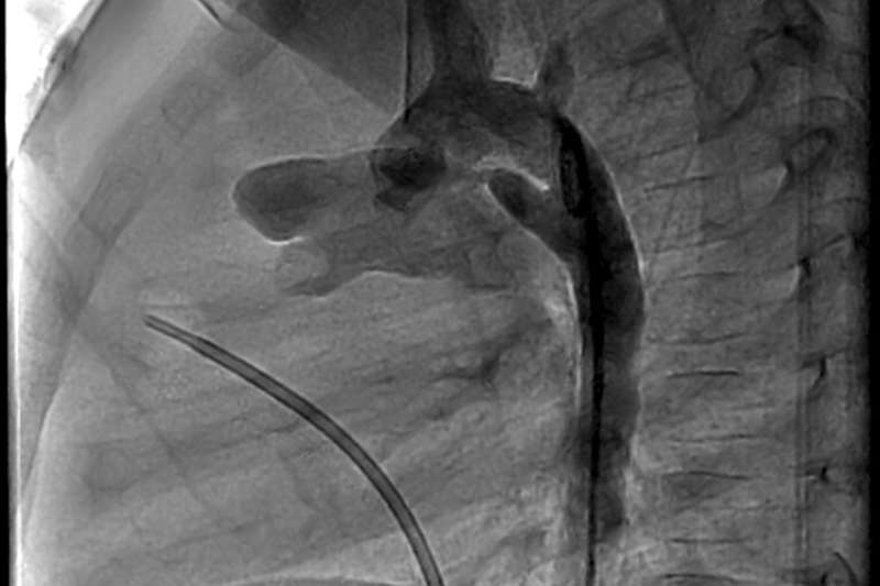
Research reveals optimal approach for patent ductus arteriosus

An X-ray shows a patent ductus arteriosus (File photo)

Doctors have long debated whether very premature babies with patent ductus arteriosus (PDA)—a cardiac condition where a blood vessel that normally closes shortly after birth remains open, stressing the heart and lungs—should be immediately treated with medication to close the vessel, or whether it’s better to wait and see if the vessel closes on its own.

Compelling new research by Children’s of Alabama neonatologist Waldemar A. Carlo, M.D., reveals important answers. Along with researchers from 32 other hospitals across the United States, Carlo sought to learn whether simply monitoring infants and intervening only if needed, compared with actively treating PDA, improves survival or reduces serious lung problems.

Results of the study, published last December in JAMA, surprised even Carlo. Monitoring infants and intervening only if needed, rather than treating the PDA up front, improved survival or serious lung problems—the main outcome—with the rate of developing bronchopulmonary dysplasia (a serious chronic lung disease) essentially the same in both groups. Notably, however, babies in the watch-and-wait group were significantly more likely to survive: 4.1% of infants in the expectant management group died, compared to 9.6% of those in the medication treatment group.

Waldemar Carlo, M.D.

“I don’t think anyone was expecting to see such a large difference in mortality,” said Carlo, who’s also the Edwin M. Dixon Professor of Pediatrics and director of the Division of Neonatology at the University of Alabama at Birmingham (UAB).

PDA is the most common cardiovascular condition in preterm infants, and Children’s clinicians monitor or treat about 100 babies with the condition each year. Occurrence of PDA jumps to more than 50% for extremely premature infants such as the 482 included in the study, who were all born between 22 and 28 weeks of pregnancy.

“PDA tends to be present in the sickest premature babies with the worst outcomes, which is why there has been a lot of interest in closing the PDA,” Carlo says. “But the data have been controversial on whether these babies need treatment or not.”

A small PDA might cause no symptoms and resolve on its own. Larger ones can lead to a variety of dangerous complications, including rapid breathing or shortness of breath, poor feeding, failure to thrive, fatigue, and frequent respiratory infections. In the study, which was carried out as part of the National Institute of Child Health and Human Development Neonatal Research Network, expectant management proved superior to all medications or other approaches used in babies randomly assigned to active treatment, including acetaminophen, ibuprofen, or indomethacin as well as surgery.

“Treatment to close the patent occurred in both groups, which is mimicked in clinical practice,” Carlo said, “and none of the treatments really had a benefit. The study design was pragmatic and easy to implement. It can help clinicians in a practical way.”

The trial reinforced how neonatologists at Children’s have long managed PDA, Carlo noted, but participating in such a high-profile, comprehensive effort benefits everyone involved.

“Our patients can take advantage of state-of-the-art care through randomized, clinical trials and innovative inventions. The trial is funded by the government, and the government benefits because we find out what are the right treatments in these patients,” Carlo said. “It’s a win-win situation.”

Neonatology

Studying magnesium sulfate in very preterm babies

Research shows magnesium sulfate does not improve neurodevelopmental outcomes in very preterm babies. (Stock photo)

By Anna Jones

Research led by Children’s of Alabama and the University of Alabama at Birmingham (UAB) shows that magnesium sulfate does not improve neurodevelopmental outcomes in babies born very preterm between 22 and 24 weeks gestation.

Babies born during 22-24 weeks of gestation have a higher risk of neurodevelopmental impairment. While prior studies have found that magnesium sulfate helps prevent the development of cerebral palsy for babies born at later gestational ages between 24 and 32 weeks, this observational study led by Margaret Page, M.D., and Ashley Battarbee, M.D., in the UAB Department of Obstetrics and Gynecology, along with colleagues in the Department of Pediatrics, is one of the few studies of its kind to look at the use of magnesium sulfate for babies born at 22-24 weeks of gestation.

“This multicenter study led by Dr. Page and colleagues at UAB is important as it is the largest study designed to assess the two-year outcomes of infants born extremely preterm exposed to antenatal magnesium sulfate and/or steroids, two common treatments given before delivery,” said Wally Carlo, M.D., co-director of Neonatology at UAB and Children’s of Alabama and the Neonatal Research Network.

Battarbee says that OBGYNs cannot always stop a preterm birth, so they oftentimes need to shift their focus to how they can best prepare the baby to be born early.

“We consider interventions such as administering magnesium sulfate or antenatal corticosteroids to the pregnant patient to help improve outcomes for their baby after birth,” Battarbee said. “While the American College of Obstetricians and Gynecologists (ACOG) recently updated their guidance on corticosteroids for babies at 22 weeks, we did not have updated guidance on magnesium sulfate and wanted to explore this treatment further.”

The results of the recent study reinforce current national guidelines recommended by ACOG and the Society of Maternal-Fetal Medicine that do not recommend giving magnesium sulfate at 22 weeks and instead prioritizing antenatal corticosteroids to improve neurodevelopmental outcomes. Unlike magnesium, steroids have demonstrated benefit for infants born at very early gestational ages and remain the primary intervention when an early delivery is anticipated.

“There is not a lot of guidance on the use of magnesium sulfate in babies at this gestational age,” said Page, the lead investigator of the study. “Through this research, we were able to find that there was no benefit to using magnesium sulfate in addition to antenatal corticosteroids. This shows us that more treatment does not always equal better outcomes.”

Both Page and Battarbee emphasize that administering magnesium sulfate at that gestational age did not lead to any measurable increase in maternal or fetal harm in this study, but magnesium does carry known maternal risks, making it important not to use it without proven benefit. Additionally, using magnesium for neuroprotection in babies at these very early gestational ages when it is not effective may hinder future research in identifying interventions that truly make a difference.

This analysis was conducted using data from and in collaboration with the Neonatal Research Network, a large research network encompassing academic institutions across the United States funded by the Eunice Kennedy Shriver National Institute of Child Health and Human Development.

Neonatology

How Children’s Neonatologists Are Playing a Crucial Role in Complex Surgeries

Children’s neonatologists work with surgeons on many types of complex cases. (File photo)

In the Level IV Neonatal Intensive Care Unit (NICU), Children’s of Alabama cares for some of the sickest neonates in Alabama and the surrounding region. The unit, which expanded in 2025, frequently receives referrals from other hospitals for surgical and subspecialty care. Often, the babies cared for on the unit have conditions that require complex surgeries. In these cases, neonatologists work with surgeons and other specialists to manage the patient’s care. Jaw distraction is one example of a procedure they may manage.

Jaw Distraction Surgery and the Role of Neonatology

For some infants, a small or recessed jaw creates serious problems with breathing and feeding. Jaw distraction surgery—also called mandibular distraction osteogenesis—is an option that may dramatically improve outcomes for these patients. While the surgery itself is performed by craniofacial surgeons, the care before and after is deeply collaborative. Neonatologists play a central role in guiding babies and families through every step of the process.

Physicians typically consider jaw distraction surgery for young infants, often within their first month or two of life. Babies with the condition often work harder to breathe normally. They also may have difficulty feeding by mouth because the small jaw narrows the airway. If growth alone doesn’t lead to improvement, then a multidisciplinary team, including neonatologists, craniofacial surgeons, and often ear, nose, and throat (ENT) surgeons, evaluates whether jaw distraction could improve their symptoms and quality of life.

Surgeons focus on the technical aspects of lengthening the jaw, but before and after the operating room, neonatologists manage the airway, ventilation, nutrition and pain control. After the procedure, the baby returns to the NICU sedated and temporarily paralyzed. This protects the infant’s airway and allows the surgical site to heal while the jaw is gradually lengthened over several days. During this time, infants require ventilator support, careful airway management, pain control and nutritional support.

“This isn’t a procedure we recommend lightly,” Children’s of Alabama neonatologist Hannah Hightower, M.D., explained. “Protecting the airway is our number one concern for infants who undergo this surgery.” Because surgeons place hardware in the jaw, any movement could risk complications. This also means post-op babies cannot be held. This period can be especially hard on new parents who may have already begun holding and feeding their child.

As recovery progresses, collaboration becomes increasingly important. Neonatologists and surgeons work together to determine when it is safe to reduce sedation, remove the breathing tube and transition the baby off respiratory support. “Then, we begin working on the infant’s oral skills, because our ultimate goal is for the family to be able to feed their baby at home,” Hightower said. Many of these infants require tube feeding at first, but most eventually learn to feed by mouth.

Even in ideal cases, infants typically remain hospitalized for several weeks as they recover, learn to eat and prepare for discharge. Parents play a significant role as the baby’s recovery progresses, becoming actively involved in feeding and comforting their child. Success after jaw distraction is measured in practical, meaningful ways, such as improved breathing without respiratory support and the ability to take feeds by bottle.

Though the procedure can seem daunting, Hightower emphasizes its impact. Jaw distraction surgery can transform the course of an infant’s life—turning a situation marked by breathing struggles and feeding challenges into one where a baby can go home, breathe comfortably and be fed by their family. “It’s remarkable how a child who is working to breathe and unable to eat may improve and gain these skills post-surgery,” Hightower said. “Being able to send a child home doing those normal baby things is a big deal for the parents.”

Neonatology

Extended CPAP Shows Promise in Preemies

Research at Children’s and UAB shows that keeping premature infants on CPAP longer may improve lung growth.

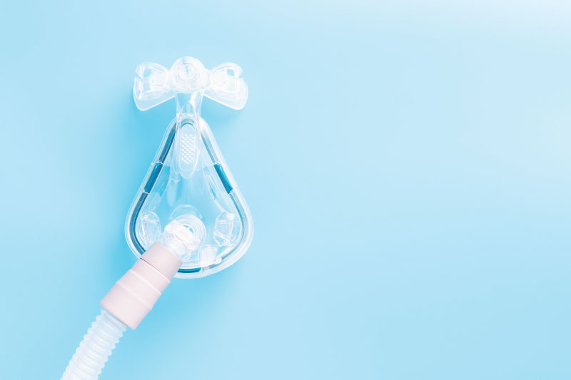
Research at Children’s of Alabama and the University of Alabama at Birmingham suggests that keeping premature babies on nasal continuous positive airway pressure (CPAP) longer than currently practiced could significantly improve respiratory outcomes, potentially changing how neonatologists approach respiratory care for the smallest patients.

Early use of CPAP is standard for preterm infants unable to breathe on their own. It helps avoid invasive ventilation and minimizes the risk of lung injury while also increasing lung volume, which could stimulate lung growth and development.

Zaki Yazdi, M.D.

Children’s neonatologist Zaki Yazdi, M.D., conducted a pilot study as part of his fellowship to see whether extending CPAP beyond traditional stopping points benefited premature infants. Yazdi’s study, published in Archives of Disease in Childhood: Fetal & Neonatal, showed that continuing CPAP reduced episodes of drops in heart rate and oxygen levels in preterm infants. These positive findings align with groundbreaking research published this year in the American Journal of Critical Care Medicine, suggesting that extending CPAP promotes lung growth in babies born prematurely.

“We know CPAP helps premature babies with respiratory distress syndrome and apnea of prematurity,” Yazdi said. “The question we were trying to answer was: When is the best time to stop CPAP? We hypothesized that if you were to continue CPAP for a 24-hour period instead of going down to nasal cannula, you would have fewer drops in your oxygen level.”

Yazdi and the Children’s neonatology team, including neonatologist Colm P. Travers, M.D., randomized 36 infants born before 34 weeks gestation to either stop CPAP when they met Children’s traditional criteria (minimal oxygen support and few apnea episodes) or continue for an additional 24 hours. The primary outcomes were oxygen levels and other vital sign changes.

Colm Travers, M.D.

Babies who remained on CPAP an additional 24 hours experienced significantly fewer episodes of intermittent hypoxemia—defined as oxygen saturation below 85% for 10 seconds or longer—compared to those transitioned to low-flow nasal cannula. The CPAP group also had fewer heart rate drops and spent less time with low oxygen saturations.

“Even though all the markers we traditionally look at say this baby should be ready to come off CPAP, perhaps there are some more subtle things that we wouldn’t normally pick up on that suggest staying on CPAP could be helpful,” Yazdi said.

Extended CPAP isn’t without risks. Prolonged treatment can delay oral feeding, since many hospitals avoid feeding babies while on CPAP. There’s also risk of nasal breakdown from the CPAP mask interface, increased costs, and potential complications like feeding intolerance from swallowing air. However, Yazdi’s study found no negative effects during the 24-hour extension period.

The team has now received funding from the National Institute of Child Health and Human Development’s Neonatal Research Network to perform a much larger study examining extended CPAP’s effects on lung development. The multicenter, randomized clinical trial—led by UAB—will involve approximately 860 babies. Unlike Yazdi’s 24-hour study, neonates randomized to the longer CPAP arm will remain on the support for at least two weeks or until they are 34 weeks post-menstrual age. The children will then be followed for two years to assess lung function development and long-term respiratory outcomes, making it the largest controlled trial of extended CPAP to date. 

Already, Yazdi said, he and other neonatologists have noticed “a little bit of creep” toward keeping babies on CPAP longer at Children’s and other institutions.

“I don’t think we’re ready to say that this is definitely the best way to go yet,” Travers said. “But preliminary data that’s very promising suggests we need to do this larger trial to see if there’s any long-term benefit.”

“This could redefine what the standard of care could be,” Yazdi said.

Neonatology

A closer look at the effects of chorioamnionitis on premature babies

The majority of preterm births stem from chorioamnionitis. (Stock photo)

The vast majority of preterm births—especially “micro-preemies” born at 22 or 23 weeks’ gestation—stem from a single cause: chorioamnionitis, an inflammation of the placenta and membranes surrounding the fetus. But Children’s of Alabama neonatologist Viral Jain, M.D., is on a mission to determine why this insidious condition occurs, the ways it affects babies’ health, and how to stop it.

Occurring in an estimated 1% to 5% of births in the United States, chorioamnionitis—often shortened to chorio—can be hard to spot. It’s typically diagnosed using clinical signs of inflammation such as fever or elevated heart rates in either the mother or the baby. But chorio often eludes clinical diagnosis, silently causing damage to the placenta and triggering preterm birth, says Jain, also an assistant professor in the Division of Neonatology at the University of Alabama at Birmingham (UAB).

Viral Jain, M.D.

“It’s a huge reason why neonatology exists, as such,” he explained. “It’s the body’s reaction when there’s inflammation to deliver the baby preterm, and all the complications that come with a preterm baby are due to chorio. In addition, the inflammation also causes direct damage to the developing organs of the baby.”

Some of the extensive research conducted on chorio has focused on its causes, which may include infection, environmental chemicals, smoking and bleeding. But scientists still have a poor understanding of why it happens, Jain notes, as well as how to catch it early enough to stop premature delivery.

Much of Jain’s research has delved into chorio’s potential health implications for babies once they’re born—and the effects can be devastating. One of his studies shows that the incidence of cerebral palsy is far higher in infants born when chorio progresses to such a severe extent it becomes funisitis, or inflammation of the umbilical cord. Jain’s findings have been somewhat controversial, he acknowledges, since cerebral palsy is already known to affect more preterm infants than those born after full-term pregnancies.

“We chose the most severe chorio babies for the study to clearly show that it affects cerebral palsy development,” Jain said. “We found that it’s about 50-50—so half the risk of cerebral palsy was from being born pre-term due to chorio, and half was the direct injury coming from inflammation to the developing brain.”

To help predict the cerebral palsy risk of these infants while they’re still in the neonatal intensive care unit (NICU)—when early intervention can more easily be planned—Jain’s research has also used MRI to look for specific markers in the brain suggesting a high risk of the disabling condition.

“We showed that chorioamnionitis insult, which started at birth, continues in these babies and that we can see those changes in the MRI and that they lead to cerebral palsy,” he said. “This means you can start early intervention on those babies to capture or reduce some of the damage.”

Another of Jain’s studies suggests that infants born early due to chorio have chronic lung damage. “It creates an immune cell dysfunction in the lung that there is continuous damage happening,” he explained. In addition to requiring longer ventilator and oxygen treatment, these babies “end up developing what we call BPD, or bronchopulmonary dysplasia, which is neonatal chronic lung disease.”

Ultimately, Jain says, his research—which has been funded by the American Heart Association and National Institutes of Health—seeks to learn how chorio propagates so doctors can impede its damage.

“The goal is to find out what treatment we can give so when it’s just mild we can stop the progression and it won’t become full-blown chorio and end up delivering the baby preterm,” he said. “If we can do that, we can prevent a lot of organ damage to the lung or brain.”  

For more information on Jain’s work on chorio, listen to this episode of the Children’s of Alabama PedsCast podcast.

Neonatology

Using mitochondrial genetics to predict BPD

Researchers at Children’s and UAB are exploring how mitochondrial function may help predict BPD risk.

Bronchopulmonary dysplasia (BPD), a chronic lung condition affecting some extremely preterm infants, continues to be a significant clinical challenge in neonatology. While often lifesaving, supplemental oxygen can be a key contributor to long-term pulmonary complications in this vulnerable population. At Children’s of Alabama and the University of Alabama at Birmingham (UAB), researchers are exploring how mitochondrial function may hold the key to understanding and preventing BPD.

Jegen Kandasamy, M.D., an associate professor in the Division of Neonatology at UAB, leads a multidisciplinary team supported by a research grant dedicated to studying mitochondrial dysfunction in BPD. The research centers on individual differences in how mitochondrial DNA (mtDNA) haplogroups—genetic variations inherited maternally and varying by ethnicity—may influence an infant’s susceptibility to lung injury from oxygen exposure, particularly hyperoxia.

“Hyperoxia is a double-edged sword,” Kandasamy said. “It’s essential for survival, yet it introduces oxidative stress that preterm lungs are poorly equipped to handle. Our research is aimed at understanding how mitochondrial genetics impact that response.”

Using collected blood samples and clinical data from preterm infants, Kandasamy’s team is working to identify mtDNA haplogroups associated with higher BPD risk. The goal is to develop precise, genetically informed risk profiles that allow for early intervention. Hopefully, this will improve outcomes while addressing racial disparities in BPD prevalence and severity.

An especially promising area of research is platelet bioenergetics. By measuring how platelets utilize mitochondrial energy, the researchers hope to identify specific biomarkers that reflect systemic mitochondrial health and may help predict BPD risk. “Platelets are easy to access and give us a real-time snapshot of mitochondrial function without invasive procedures,” Kandasamy noted.

The team is also studying mitophagy, the elimination of damaged mitochondria through autophagy, and its role in lung development. Emerging evidence suggests that impaired mitophagy contributes to persistent mitochondrial dysfunction, exacerbating lung injury in preterm infants. As a result of this new evidence, the group is also evaluating the potential of thyroid hormone supplementation as a therapeutic strategy to restore mitochondrial function and mitigate lung damage.

By integrating clinical data with mouse models, the UAB team is uniquely positioned to investigate both the mechanistic underpinnings of BPD and potential interventions. The collaborative effort spans neonatology, mitochondrial biology and pediatric pulmonology, creating a comprehensive research environment.

“Our ultimate aim is to shift the paradigm from reactive to predictive personalized neonatal care,” Kandasamy said. “Understanding how mitochondrial genetics intersect with environmental exposures can help us identify at-risk infants earlier and intervene more effectively.”

Neonatology

Pandemic Practices and Neonatal Health Issues

A study led by Children’s researchers shows the link between pandemic health behaviors and neonatal health issues.

By Hannah Echols, UAB

Studies show that social distancing and other public health measures during the COVID-19 pandemic effectively reduced the spread of the deadly virus. However, they had unanticipated effects such as reduced health care accessibility and utilization, especially in high-risk populations.

Researchers at Children’s of Alabama and the University of Alabama at Birmingham (UAB) evaluated potential effects of pandemic-related behavior changes on neonatal mortality and preterm birth rates. A correlation was found between the social distancing index, a measure of overall social distancing behaviors observed, and higher rates of neonatal and early neonatal mortality, as well as preterm birth, when assessed with a lag period. Results were published in July in JAMA Network Open.

“COVID-19 affected the health care systems globally, and many lives were lost; it is important to learn from this experience to prepare better for possible future health crises,” said Vivek Shukla, M.D., assistant professor in the UAB Division of Neonatology and lead author of the study. “We need to understand how changes in health behavior affected outcomes, whether people had limited access to care or healthy habits were altered.”

Maternal pregnancy complications increase the risk of preterm delivery and neonatal morbidity. These complications are a major contributor toward neonatal mortality. During the COVID-19 pandemic, pregnant women encountered substantial obstacles in accessing health care. 

According to the American Medical Association, 81 percent of physicians were providing fewer in-person visits when surveyed in July and August of 2020 than pre-pandemic and the average in-person visits fell from 95 to 57 per week.

“The observed correlations may be due to changes in health care access during periods of increased social distancing, such as fewer prenatal visits,” said Rachel Sinkey, M.D., associate professor in the UAB Division of Maternal-Fetal Medicine and co-author of the study. “These appointments are important to catch and address complications that could be life-threatening to both mom and baby.”

Defining the trend

The population-based study used data from the Centers for Disease Control and Prevention’s National Center for Health Statistics. The team evaluated neonatal mortality and preterm birth rates from 2016 to 2019 and compared them to 2020 rates. In unadjusted comparison, the rates appeared to be lower. When adjusted for a declining trend observed in the 2016-2019 period using an Auto Regressive Integrated Moving Average model, the rates were not significantly different.

Shukla further analyzed the correlation between the Social Distance Index—which indicated overall population mobility during the pandemic—and neonatal mortality and preterm birth rates in 2020. On a first look, there was no significant correlation; but when a lag period was added, higher SDI was associated with higher neonatal mortality rates with a two-month delay and with higher preterm birth rates with a one-month delay.

“With these in-depth analyses, we could account for the effect of a potential delay, or lag time, receiving access to care could have on the mortality and preterm birth rates,” Shukla said.

“The results indicate a need for more in-depth studies on the unintended effects of pandemic-related health behavior changes,” he added. “Conducting additional studies is an important step for providers and public health experts to better prepare in case there is a next public health crisis.”

Neonatology, Pulmonology

Improving lung function for COPD and BPD patients

A study led by Children’s researchers shows that inhalation of live Lactobacilli reduces inflammatory markers in BPD and COPD.

By Jeff Hansen, UAB

In preclinical models, the inhalation of a mixture of living Lactobacilli bacteria attenuated pulmonary inflammation and improved lung function and structure for the chronic lung diseases bronchopulmonary dysplasia (BPD) and chronic obstructive pulmonary disease (COPD).

This study, published in the journal Nature Communications, determined the mechanism of this live biotherapeutic product—a powder mixture of living Lactobacilli bacteria—to reduce neutrophilic inflammation and reduce a broad swath of inflammatory markers in BPD and COPD, says Charitharth Vivek Lal, M.D., a neonatologist at Children’s of Alabama and the University of Alabama at Birmingham (UAB). Lal co-led the research with Amit Gaggar, M.D., Ph.D., a UAB pulmonologist.

Their findings “provide a paradigm for the progression of structural lung disease,” Lal said, because they identify the Lactobacilli as critical to regulating lung protease activity that is linked to the destruction caused by matrikine generation, extracellular matrix turnover and chronic neutrophilic inflammation that damages air sacs in the lungs. 

A possible protective role for Lactobacilli in the lung and the possible use of Lactobacilli to treat chronic lung disease had its foundation in 2016 when Lal and UAB colleagues discovered that the airways of infants with severe bronchopulmonary dysplasia had decreased numbers of Lactobacilli, increased numbers of proteobacteria and increased concentrations of proteobacterial endotoxin. In this latest study, the UAB researchers provided a mechanism of action for the Lactobacilli treatment to decrease downstream disease development and showed safety and effectiveness of the live biotherapeutic treatment in a mouse pup model for BPD and three mouse models of COPD. 

Bronchopulmonary dysplasia develops in some extremely premature infants after damage induced by high oxygen tension or mechanical ventilation needed to keep them alive. COPD occurs in older people, especially smokers, and kills about 130,000 Americans a year and about 3 million more worldwide.

“Inhaled live biotherapeutic products show promise in addressing common pathways of disease progression that in the future can be targeted at a variety of lung diseases,” Lal said. “Preclinical animal data is suggestive, and safety of the potential drug in humans will be tested in a forthcoming clinical trial. Human adult safety data in COPD will help de-risk the pathway to approval for use of the drug in bronchopulmonary disease infants.”

The UAB researchers hypothesized that mouse models of BPD would show heightened levels of acetylated proline-glycine-proline, or Ac-PGP, an extracellular matrix-derived peptide, as had been seen in premature infants with BPD.

This was demonstrated in BPD mouse models, and gain- or loss-of-function studies showed the impact of Ac-PGP. Intranasal instillation of Ac-PGP increased neutrophilic inflammation and lung degradation. When an inhibitor of Ac-PGP was given with the Ac-PGP, markers of neutrophilic inflammation decreased and lung structure improved.

Researchers then showed that a proprietary Lactobacilli blend of L. planatarum, L. acidophilus and L. rhamnosus performed best in synergy to reduce the inflammatory proteinase MMP-9, which helps release the Ac-PGP from extracellular matrix. Furthermore, supernatant from Lactobacilli growth medium also reduced MMP-9 at a similar magnitude as live Lactobacilli bacteria. 

A key finding was that L(+) lactic acid, which is produced in Lactobacilli growth medium supernatant, reduced MMP-9 in vitro, showing an important role for this lactic acid as an anti-inflammatory molecule. Researchers found that live Lactobacilli in the lungs provided an ongoing, sustained release of L(+) lactic acid in a controlled and well-tolerated manner.

A major technological advance reported in the study was creating the inhaled Lactobacilli powder through particle engineering—particles small enough to reach deep into the lungs while preserving viable bacteria. This live biotherapeutic product was then tested in the BPD and COPD models. In the COPD mouse models, the blend successfully reduced inflammation in the lung microenvironment whether treated concurrently or post-injury, showing anti-inflammatory effects, decrease of several pro-inflammatory markers and elevation of the anti-inflammatory marker IgA. 

An interesting finding was the favorable performance of the live biotherapeutic product. It reduced MMP-9 and other pro-inflammatory cytokines as effectively as—or in some cases better than—fluticasone furoate, a United States Food and Drug Administration-approved inhaled corticosteroid found in COPD combination therapies. 

Safety and biodistribution studies in one of the COPD mouse models showed that inhalation of the bacterial powder did not initiate adverse reactions or disease, and the Lactobacilli did not translocate to distal tissues or accumulate in the lungs.

Neonatology

Innovative Lung Testing for Preemies

A device developed by Children’s of Alabama neonatologists is showing promise at measuring lung function in premature babies.

One of the most important parameters of a premature baby’s health status is respiratory health. How well are their lungs working? What long-term respiratory complications might occur from the premature birth? Yet assessing lung function in these fragile newborns has long been a complex and invasive process.

“Right now, we diagnose lung disease based on whether a baby needs oxygen or not,” Children’s of Alabama neonatologist Colm P. Travers, M.D., said. “But we don’t know how severe their lung disease is and what type of lung disease they have.”

Measuring lung function in children and adults is as simple as having them blow hard into a plastic tube. Pulmonary function testing in neonates, however, requires complicated equipment as well as sedation or anesthesia, limiting its use outside of research studies.

Colm Travers, M.D.

So, Travers and his team have brought a decades-old technology used in adults called forced oscillation technique (FOT) to the NICUs at the University of Alabama at Birmingham (UAB) and Children’s. They worked with the device manufacturer to develop a machine designed for small babies. It sends sound waves into the lungs while the infant is sleeping and breathing naturally. The sound waves then bounce off the lungs, providing information on stiffness and resistance in the airways. It can be performed in less than 30 seconds, non-invasively, without sedation and repeated over time. It typically takes just a few minutes to set up and can be used soon after birth.

“It’s well suited to neonates because it doesn’t need any effort on behalf of the baby,” Travers said. “Oscillometry is also able to tell you the severity and the type of lung disease a baby has.” In addition, he said, it’s an ideal tool to use in research because of its simplicity and accuracy.

The Children’s team has already used the device in more than 600 neonates. They published the first results of their work in 2020, which demonstrated the feasibility of using oscillometry in healthy newborns and those with respiratory disorders. Now, they’re in the midst of a larger study funded by the National Heart, Lung, and Blood Institute involving 550 babies with the goal of establishing normative data for infant lung function and tracking outcomes, such as asthma and wheezing, in early childhood.

“We’re also using it in our NICU to see whether babies respond to certain medications,” Travers said. This approach could help doctors quickly determine if a treatment is effective or if they need to pursue alternative options.

The use of FOT at Children’s is still only in the research phase. Travers says he and his team are still following the infants until they are 2 years old. “In the future, we hope to see the infants back around the time they are starting school so we can see how they are doing in terms of exercise capacity and lung function,” he said.

Eventually, Travers hopes FOT will become standard of care not only in his NICU, but in hospitals everywhere.