

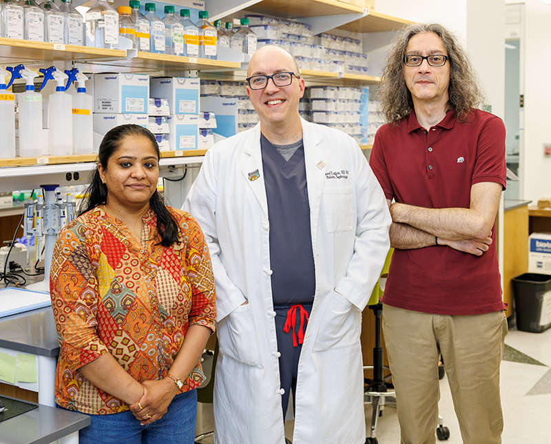
In the UAB Spatial Core Lab, researchers are using spatial transcriptomics to examine specific regions within tissue samples.
A breakthrough technology that allows doctors to study the precise regions of a kidney transplant that are involved in rejection could transform how doctors learn about kidney transplant biology, potentially leading to new diagnostic tests or treatments for children.
The technology, called spatial transcriptomics, is a leading-edge technique that lets researchers see which genes or proteins are active in very specific regions of tissue samples, such as those from a kidney biopsy. Unlike traditional methods that study tissue samples as a whole without regard for the location of important signals from the tissue, this technique allows researchers to examine custom-shaped regions of interest containing just a few cells in their natural environment.
Think of it as having a detailed map that shows not just what’s happening in a city, but precisely in which neighborhoods each activity occurs.
“There’s been decades now of data showing that gene expression patterns coming from a transplant are a little bit more sensitive for problems coming from a kidney transplant,” said Michael Seifert, M.D., director of the University of Alabama at Birmingham (UAB) Spatial Core and medical director of pediatric renal transplantation at Children’s of Alabama. “The problem is that we’ve never exactly known where those signals are coming from. Are they coming from cells in the kidney that we care about or are they coming from cells in the kidney that may not be as relevant?”
For instance, signals from immune system cells would be extremely relevant, he said, but could be distinct from those coming from the endothelial cells lining blood vessels.
With this technique, “we can look at a picture of a kidney biopsy on our instrument screen and take your mouse and draw a shape around it, and it will profile everything in that shape while ignoring everything else around it,” he said. It can even profile a certain cell type within the shape.
The technology itself isn’t destined for routine clinical use, Seifert said. “I can’t foresee a scenario where I would do a biopsy and then use spatial transcriptomics to make a diagnosis, because it’s a very labor-intensive, time-intensive and cost-intensive technique.”
Instead, he said, “my hope is that this will allow us to have a deeper understanding of the processes involved in transplants doing well but also transplants doing poorly. That will help us design better management programs, whether that’s using existing medicines in different ways or designing new medicines that can be more targeted and more effective than what we currently have available.”
Understanding exactly which parts of the kidney are affected by rejection also opens the door to personalized transplant care.
“Every cell in the kidney behaves differently depending on where it sits,” Seifert said. “This technology lets us uncover the heterogeneity—that is, the differences—within the tissue,” including if the problem lies in the blood vessels or the tubules or the parts of the kidney that generates urine. “I hope that’ll allow us to understand the signals that vary from person to person so we can really apply that more personalized technique.”
Thus, rather than treating all kidney transplant patients the same way, doctors could tailor anti-rejection treatments based on what is happening in an individual child’s kidney. This would, however, require advances in the spatial transcriptomics technology to make it faster and less expensive.
Spatial biology is not limited to the study of kidney transplant diseases. Seifert and his team in the UAB Spatial Core are working with specialists in other disciplines throughout Children’s and UAB, including ophthalmology, oncology and pulmonology. “We’re open to collaborating with any investigator with a good question that spatial biology can answer,” he said.
In fact, he sees spatial biology as an important technique for understanding all diseases in children. “I think what’s come out of this is an appreciation that the spatial context is incredibly important in so many of the diseases that we study.”
