All Posts By

childrensal

Neurology & Neurosurgery

Children’s of Alabama Offering New Gene Therapy for Patients With DMD

A new treatment offers the hope of longer, better life for DMD patients. (Stock photo)

In January 2025, Children’s of Alabama, for the first time, treated a patient with Duchenne muscular dystrophy (DMD) using a new gene therapy offered by only a few academic hospital facilities in the nation. The milestone followed a lengthy approval process and marked a new opportunity for patient success and scientific progress. Though not a cure, the treatment represents the hope of a longer life for these patients. For researchers, it will contribute to greater learning about the potential of this new treatment.

What is DMD?

While it is considered a rare disease, DMD is the most common form of muscular dystrophy, affecting one in every 5,000 males born in the United States. Patients experience progressive muscle degeneration, starting with proximal muscles and expanding to the limbs over time. They have trouble with many physical activities such as jumping, running and walking, and lose the ability to walk over time. The disease is fatal, and most patients don’t live past their late 20s. DMD has no cure; treatment focuses on extending the patient’s life by slowing down the disease’s progression.

According to the Muscular Dystrophy Association, symptoms of DMD can begin as early as ages 2-3 years, but in Alabama, where DMD is not yet part of newborn screening, many boys are not diagnosed until ages 4-6. That, says Children’s neuromuscular nurse practitioner Samantha Weaver, DNP, CRNP, is when patients begin to experience a steady decline. 

Treatment

Since the 1990s, physicians have prolonged the lives of patients with DMD using corticosteroids, whose anti-inflammatory properties can slow down the disease’s progression by about three years. Gene therapy, however, represents a new treatment aimed at restoring the function of the causative gene, DYSTROPHIN. The U.S. Food and Drug Administration originally approved it in 2023 for use in patients ages 4-5. In 2024, the agency extended that approval to all patients 4 years and older.

In this treatment, the transgene (a micro-DYSTROPHIN synthetic gene) is packaged within a viral capsid—a virus not intended to harm the patient that can hold the genetic material. In essence, physicians are “giving back the missing genetic information to the muscle tissue,” Children’s neurologist Michael Lopez, M.D., Ph.D., said. Unlike any other option, he noted, gene therapy treats the root cause of DMD.

“Now that we’re starting to get these really breakthrough therapies, they’re fulfilling on the promise that we all were searching for, which is that we could get closer to making this disease really better,” he said.

For the patient, improvements don’t happen overnight. That’s not how gene therapy works, Lopez says. “What we hope is that over many years, we’ll see a slow progression of the disease that is beyond what we would get with just treatment with corticosteroids alone,” he explained. “And I think that added benefit is something that’s going to be more of a long-term improvement.”

So, what can the parents of each patient treated with gene therapy hope to see? Ideally, in the short-term, their child will be more active. “I hope that our children can have more of a shot at more play and more jumping and more climbing and all of those things in the future,” said Erin McLeod, M.D., a pediatric neuromuscular neurologist at Children’s. In the long-term, the hope is that they’ll have a longer life.

Evidence supports the treatment’s efficacy. The clinical trials show that gene therapy is being delivered to patient’s muscles, and while the motor assessments haven’t shown clear evidence of clinically observable benefits, the data has trended toward improvement. Lopez says in other, more-recent studies, treated patients are starting to show improvements compared to those not receiving gene therapy. “The MRIs of the muscles themselves look a little bit healthier in some of these patients,” he said. “There’s less evidence of disease in that.”

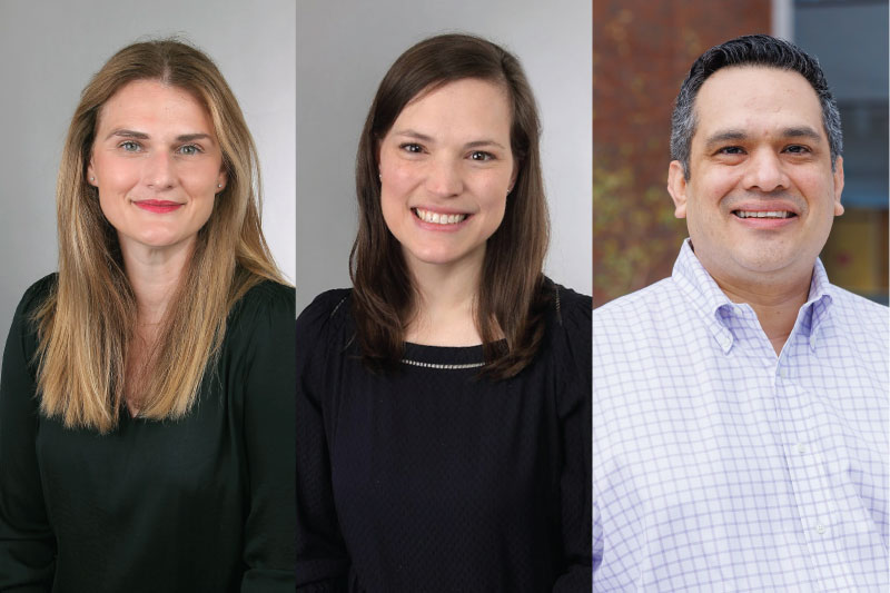
From left: Samantha Weaver, DNP, CRNP; Erin McLeod, M.D.; Michael Lopez, M.D., Ph.D.

Finding the Right Fit

Gene therapy, however, is not the right fit for every patient. To determine candidates, Children’s looks at age, underlying disease and disease progression, Weaver said. They also consider the patient’s overall health and risk for infectious diseases. “It’s an extensive process,” she explained.

With all gene therapies, safety must be prioritized. The treatment can produce a significant immune response that can even prove life-threatening to patients with more advanced stages of the disease. Liver injury is also a major concern. Thus, Weaver says the team must ensure the patient has no antibodies that will reject the virus. “These are important steps to make sure the patient will have the best outcome,” she said.

Because of these considerations, only a small percentage of patients are ideal for the treatment.

Why Offer it at Children’s?

In Alabama, Children’s is the only hospital that offers gene therapy for patients with DMD. Making it available made sense—the hospital already treats spinal muscular atrophy (SMA) patients with gene therapy. Brad Troxler, M.D., and Shelley Coskery, CRNP, led the way on that, Lopez said, and “built in a lot of the infrastructure that we needed to be able to start doing gene therapies.”

“That really has put us out in front of the field with the experience to deliver these high-cost and novel leading-edge treatments,” he added.

Challenges

Cost was one of many challenges for the team as they sought approval to implement this multimillion-dollar therapy. To that end, they involved hospital administration and a pharmacoeconomics committee in the process. But, as Weaver pointed out, the process involved many more steps including determining who would write a protocol to ensure patient safety. They also had to build a larger team, which ultimately included hepatologists, pulmonologists, cardiologists, physical therapists and social workers.

Obstacles persist, even as Children’s offers the treatment. “A high cost remains a big challenge,” Lopez said. “And so we’ve been fortunate to be able to provide these treatments because we’ve gotten support from the insurers, so far.” But not every insurer is the same, he noted, and some may be slow to cover or even decline to cover the treatment.

What the Future Holds

So far, Children’s has dosed only one patient—out of roughly 100 that it follows—with the new gene therapy. While few will be candidates for the treatment, the team hopes to dose more in the future, as long as “the risk is appropriate and the benefit is continuing to be demonstrated,” Lopez said. The team also expects more advancements, which may make it possible for others—especially those with more severe cases—to receive the treatment.

“This first approved treatment for Duchenne that is a gene therapy is just the beginning, and there are going to be more down the road,” Lopez added. “There are certainly some that are in clinical trials now. So I think we’re right to be optimistic in that we’re starting to really push the treatment of Duchenne in a way that’s going to give us lots of options that weren’t there before.”

And as Children’s continues to offer the treatment, they’ll contribute to the scientific community’s information on its effectiveness, which means the team is paving the way toward greater success for the broader population of patients with DMD.

“I think everyone who is familiar with it at this point knows it is not a cure. But it is supposed to significantly slow the disease, and we are still gathering and gaining more data to that,” Weaver said. “So we’re very excited to be part of that process.”

Urology

A new protocol for kidney tests in spina bifida patients

A new study shows that ultrasound is not enough to monitor kidney health in children with spina bifida. (Stock photo)

For decades, doctors have relied heavily on ultrasound scans to monitor kidney health in children with spina bifida, the most common permanently disabling birth defect in the U.S. People with spina bifida tend to develop end-stage renal disease up to 20 years earlier than the general population, so keeping a close watch on kidney health from a young age is important, says Children’s of Alabama pediatric urologist Stacy Tanaka, M.D. “Then if there’s a concern, it can be acted upon early and not ignored.”

Current guidelines from the Spina Bifida Association (SBA) recommend annual screening with ultrasound to look for hydronephrosis—a condition in which the urine backs up into one or more kidneys—as a sign of kidney function, and blood tests like serum creatinine, to measure overall kidney health. But with kids, Tanaka says, “the practice pattern was that a lot of people were only doing renal ultrasound.”

Now a new study from Tanaka and her Children’s colleague David Joseph, M.D., as well as other kidney experts from around the country, shows that ultrasound alone is not enough to assess kidney health. “We basically use ultrasonography as a reflection of renal function,” Joseph said, but few, if any, studies assessed its accuracy in determining renal function.

Stacy Tanaka, M.D.

The study’s genesis came during a multidisciplinary meeting in 2003 of specialists who treat children with spina bifida. “The bottom line from all disciplines at that time was that nobody was really treating this population with evidence-based care,” Joseph said.

To change that, the Centers for Disease Control and Prevention and the SBA established the National Spinal Bifida Patient Registry (NSBPR), to which 20 spina bifida clinics submit data to help develop evidence-based care. In addition, nine clinics established a urologic protocol to manage and preserve initial renal function in young children with spina bifida (UMPIRE). The NSBPR and UMPIRE provided the data set Joseph and Tanaka used to determine the effectiveness of renal ultrasound vs. blood test to assess renal function.

The two registries included data on 2,500 children ages 1-18 with myelomeningocele, the most severe form of spina bifida. All had had an ultrasound and blood test within six months to determine estimated glomerular filtration rate (eGFR), a marker of kidney health.

The results were striking: ultrasound-based detection of hydronephrosis had only about a 25% sensitivity for identifying children with signs of chronic kidney disease in the UMPIRE study and 24% in the NSBPR cohort. That means kidney damage in three out of four children was going undetected. The poor sensitivity held even when researchers looked only at severe hydronephrosis, which had an even worse sensitivity rate–just 6% to 11%. “The renal ultrasound by itself wasn’t all that good,” Joseph said, “but that didn’t surprise us.”

David Joseph, M.D.

The findings challenge current practice and suggest that blood tests measuring kidney function should be routinely performed alongside ultrasound, not just when ultrasound results look concerning, as some clinicians practice. The team at Children’s prefers testing for cystatin C rather than creatinine because of the test’s improved and more accurate ability to obtain an eGFR.

One reason clinicians may eschew blood tests is that it involves needles, Tanaka said, which be traumatizing for children. Ultrasound, on the other hand, is noninvasive, easily available, and can be performed by technicians.

“The ultrasound is very helpful and important,” Joseph said, “but you need to recognize that it may not be telling you about renal function or injury to the kidney.” The findings have already changed practice at Children’s, where all kids with spina bifida now receive both tests during kidney health screening.

Ideally, the next study would randomize kids to either double testing or ultrasound alone, but that requires significant funding, particularly since the children would need to be followed for years.

This study was conducted with very little financial support, Tanaka said. “It represents a labor of love for everyone at all nine UMPRIE centers who have been involved in this project,” Joseph added.

Nephrology

Reducing hypertension numbers in children—and adults

The Children’s of Alabama Hypertension Clinic aims to help children in the short-term and long-term. (Stock photo)

Successfully stemming rising rates of cardiovascular disease in adults needs to start in childhood. But with mounting numbers of children and adolescents developing high blood pressure—a trend driven largely by skyrocketing obesity rates—this objective is getting harder to achieve. Enter the Pediatric Hypertension Program at Children’s of Alabama, which, with its steady growth, seeks to break the cycle.  

The Hypertension Clinic, which operates three half-days each week, now sees about 45 patients weekly, a 10-fold increase from 14 years ago, says Daniel Feig, M.D., Ph.D., director of the Division of Pediatric Nephrology at Children’s, who was recruited in 2011 to oversee the clinic’s development and expansion.

High blood pressure—defined in adults and children 13 years and older as a reading of 130/80 mm Hg or higher—is relatively unusual in healthy young patients, affecting 2-3% of typical children and adolescents. (For younger children, the definition of hypertension is a statistical one, based on greater than 95th percentile for age, sex and height.) But children with obesity—who account for nearly 20% of all Americans under 18—have a 20-30% rate of hypertension, says Feig, also the Margaret Porter Professor of Pediatrics at University of Alabama at Birmingham (UAB).

Daniel Feig, M.D.

“There’s a fairly large number of kids with high blood pressure, and one of the major concerns is how we can impact the long-term epidemic that results in cardiovascular disease in adults,” he said. “Controlling hypertension in adults hasn’t gone very well—only about half of those diagnosed have even remotely effective control. This impacts their cardiovascular disease and stroke risk.”

“The only way we can get this under control is by prevention,” Feig added. “If we can make an impact in children and adolescents, we can push back cardiovascular disease in adults.”

Drawing patients from across Alabama as well as some from eastern Mississippi and western Georgia, the Children’s Hypertension Program provides ongoing care for about 2,200 children. But this “catchment area” is likely home to about 70,000 young people with high blood pressure who are undiagnosed. Part of the problem is that many pediatricians aren’t comfortable diagnosing or treating the condition, Feig explains.

When patients come to Children’s, they’re often set up with ambulatory blood pressure monitoring equipment they wear for 24 to 48 hours to measure blood pressure a few times each hour while doing normal activities. The technology enables Children’s clinicians to tease out who actually has hypertension and not blood pressure spikes resulting from factors such as exertion, nervousness or pain.

Once diagnosed, Feig and pediatric nurse practitioner Jessica Edmondson collaborate with dietitians, pharmacists, social workers and others at Children’s to ensure patients benefit from a multidisciplinary approach to treatment. Ultimately, they’re trying to prevent both short- and long-term health implications resulting from hypertension, including heart thickening, retinal damage and even impairments in cognitive function.

It’s a daunting task, Feig acknowledges. “Anything we can do to reduce the numbers right now has a domino effect that reduces long-term target organ damage and long-term cardiovascular risk,” he said. “We’re not at a point where we can reverse the trajectory in 70,000 undiagnosed kids in Alabama, but we can positively impact a good number of kids, improve their health and quality of life, and gain the opportunity to gradually access more and more of them.”

Neonatology

Extended CPAP Shows Promise in Preemies

Research at Children’s and UAB shows that keeping premature infants on CPAP longer may improve lung growth.

Research at Children’s of Alabama and the University of Alabama at Birmingham suggests that keeping premature babies on nasal continuous positive airway pressure (CPAP) longer than currently practiced could significantly improve respiratory outcomes, potentially changing how neonatologists approach respiratory care for the smallest patients.

Early use of CPAP is standard for preterm infants unable to breathe on their own. It helps avoid invasive ventilation and minimizes the risk of lung injury while also increasing lung volume, which could stimulate lung growth and development.

Zaki Yazdi, M.D.

Children’s neonatologist Zaki Yazdi, M.D., conducted a pilot study as part of his fellowship to see whether extending CPAP beyond traditional stopping points benefited premature infants. Yazdi’s study, published in Archives of Disease in Childhood: Fetal & Neonatal, showed that continuing CPAP reduced episodes of drops in heart rate and oxygen levels in preterm infants. These positive findings align with groundbreaking research published this year in the American Journal of Critical Care Medicine, suggesting that extending CPAP promotes lung growth in babies born prematurely.

“We know CPAP helps premature babies with respiratory distress syndrome and apnea of prematurity,” Yazdi said. “The question we were trying to answer was: When is the best time to stop CPAP? We hypothesized that if you were to continue CPAP for a 24-hour period instead of going down to nasal cannula, you would have fewer drops in your oxygen level.”

Yazdi and the Children’s neonatology team, including neonatologist Colm P. Travers, M.D., randomized 36 infants born before 34 weeks gestation to either stop CPAP when they met Children’s traditional criteria (minimal oxygen support and few apnea episodes) or continue for an additional 24 hours. The primary outcomes were oxygen levels and other vital sign changes.

Colm Travers, M.D.

Babies who remained on CPAP an additional 24 hours experienced significantly fewer episodes of intermittent hypoxemia—defined as oxygen saturation below 85% for 10 seconds or longer—compared to those transitioned to low-flow nasal cannula. The CPAP group also had fewer heart rate drops and spent less time with low oxygen saturations.

“Even though all the markers we traditionally look at say this baby should be ready to come off CPAP, perhaps there are some more subtle things that we wouldn’t normally pick up on that suggest staying on CPAP could be helpful,” Yazdi said.

Extended CPAP isn’t without risks. Prolonged treatment can delay oral feeding, since many hospitals avoid feeding babies while on CPAP. There’s also risk of nasal breakdown from the CPAP mask interface, increased costs, and potential complications like feeding intolerance from swallowing air. However, Yazdi’s study found no negative effects during the 24-hour extension period.

The team has now received funding from the National Institute of Child Health and Human Development’s Neonatal Research Network to perform a much larger study examining extended CPAP’s effects on lung development. The multicenter, randomized clinical trial—led by UAB—will involve approximately 860 babies. Unlike Yazdi’s 24-hour study, neonates randomized to the longer CPAP arm will remain on the support for at least two weeks or until they are 34 weeks post-menstrual age. The children will then be followed for two years to assess lung function development and long-term respiratory outcomes, making it the largest controlled trial of extended CPAP to date. 

Already, Yazdi said, he and other neonatologists have noticed “a little bit of creep” toward keeping babies on CPAP longer at Children’s and other institutions.

“I don’t think we’re ready to say that this is definitely the best way to go yet,” Travers said. “But preliminary data that’s very promising suggests we need to do this larger trial to see if there’s any long-term benefit.”

“This could redefine what the standard of care could be,” Yazdi said.

Neonatology

Gut fungi can predict BPD, study shows

Research shows that the gut composition of fungi in the second week of life predicts the later development of BPD.

By Jeff Hansen (UAB)

Extremely preterm newborns who weigh less than 3.3 pounds have immature lungs that often require high levels of ventilation oxygen in the hospital. This contributes to the chronic lung disease bronchopulmonary dysplasia, or BPD, the most common cause of death for these tiny infants. BPD exacts a devastating toll on the immature lung.

In one of the most extensive studies of the microorganisms in the intestines of very preterm infants, University of Alabama at Birmingham (UAB) and University of Tennessee Health Science Center researchers show that the gut composition of fungi in the second week of life predicts the later development of BPD, weeks to months before diagnosis of that disease. They analyzed gut fungi in the first true non-meconium stool produced before two weeks of life and found that the fungal intestinal microbiome—known as the mycobiome—of infants who later developed BPD differed in community diversity, composition and interconnectivity from the infants who never got BPD, as measured by the most up-to-date bioinformatic techniques. The researchers did not find significant differences in the bacterial microbiome in those first true stools.

To show causality, researchers transferred samples of the first true stool that predicts BPD or the first true stool of newborns who did not get BPD into female mice to give them a pseudo-humanized gut microflora. In a mouse model of BPD, newborn pups from those BPD dams showed an increased in the severity of lung injury compared with newborn pups from the no-BPD dams. In loss-of-function experiments, when the female mice with the BPD-predictive stool transplant were treated with an antifungal agent before birth, that inhibition of perinatal fungal colonization reduced lung injury in the newborn pups. In contrast, a gain-of-function experiment, where the perinatal fungal colonization of dams was augmented with a species of Candida fungus common in mice, amplified BPD severity in the newborn pups.

Kent Willis, M.D.

“These findings demonstrate that features of the initial intestinal fungal microbiome are associated with the later development of BPD in premature neonates and exert a microbiome-driven effect that is transferable and modifiable in mouse models,” said Children’s of Alabama neonatologist Kent Willis, M.D., who’s also an assistant professor in the UAB Department of Pediatrics Division of Neonatology. “This suggests causality, and it suggests that the gut fungi may represent a therapeutic target in newborn lung disease.”

Willis and Ajay J. Talati, M.D., University of Tennessee Health Science Center, Memphis, Tennessee, co-led the study, published in Microbiome.

“Collectively, our analyses demonstrate that the composition of the intestinal mycobiome of infants who did not develop BPD was more uniform,” Willis said. “In contrast, those who eventually developed BPD had more disparate mycobiomes. This suggests that a particular pattern of mycobiome development may be necessary to impart resistance to the development of BPD, and failure to do so in various ways is associated with disease development.”

The first defecations of newborn infants are meconium, composed from materials ingested while in the uterus. The first true stools in the second week, the ones analyzed by Willis and Talati, are digested milk. It is known, Willis and Talati say, that fungi in adults are vital members of the human microbiota; but compared to bacteria, their non-pathological and non-parasitic functions are still poorly understood, especially in newborns.

This prospective observational cohort study included newborn stool samples collected over six years from 2017-2020 in Memphis and 2021-2022 in Birmingham. The 64 very-preterm infants in the study who did not develop BPD had an average birthweight of 2.5 pounds, and the 38 very-preterm infants who did develop BPD had an average birthweight of 1.6 pounds. Only one of the 64 no-BPD infants in the study died, while six of the 38 BPD infants died.

Neonatology

A closer look at the effects of chorioamnionitis on premature babies

The majority of preterm births stem from chorioamnionitis. (Stock photo)

The vast majority of preterm births—especially “micro-preemies” born at 22 or 23 weeks’ gestation—stem from a single cause: chorioamnionitis, an inflammation of the placenta and membranes surrounding the fetus. But Children’s of Alabama neonatologist Viral Jain, M.D., is on a mission to determine why this insidious condition occurs, the ways it affects babies’ health, and how to stop it.

Occurring in an estimated 1% to 5% of births in the United States, chorioamnionitis—often shortened to chorio—can be hard to spot. It’s typically diagnosed using clinical signs of inflammation such as fever or elevated heart rates in either the mother or the baby. But chorio often eludes clinical diagnosis, silently causing damage to the placenta and triggering preterm birth, says Jain, also an assistant professor in the Division of Neonatology at the University of Alabama at Birmingham (UAB).

Viral Jain, M.D.

“It’s a huge reason why neonatology exists, as such,” he explained. “It’s the body’s reaction when there’s inflammation to deliver the baby preterm, and all the complications that come with a preterm baby are due to chorio. In addition, the inflammation also causes direct damage to the developing organs of the baby.”

Some of the extensive research conducted on chorio has focused on its causes, which may include infection, environmental chemicals, smoking and bleeding. But scientists still have a poor understanding of why it happens, Jain notes, as well as how to catch it early enough to stop premature delivery.

Much of Jain’s research has delved into chorio’s potential health implications for babies once they’re born—and the effects can be devastating. One of his studies shows that the incidence of cerebral palsy is far higher in infants born when chorio progresses to such a severe extent it becomes funisitis, or inflammation of the umbilical cord. Jain’s findings have been somewhat controversial, he acknowledges, since cerebral palsy is already known to affect more preterm infants than those born after full-term pregnancies.

“We chose the most severe chorio babies for the study to clearly show that it affects cerebral palsy development,” Jain said. “We found that it’s about 50-50—so half the risk of cerebral palsy was from being born pre-term due to chorio, and half was the direct injury coming from inflammation to the developing brain.”

To help predict the cerebral palsy risk of these infants while they’re still in the neonatal intensive care unit (NICU)—when early intervention can more easily be planned—Jain’s research has also used MRI to look for specific markers in the brain suggesting a high risk of the disabling condition.

“We showed that chorioamnionitis insult, which started at birth, continues in these babies and that we can see those changes in the MRI and that they lead to cerebral palsy,” he said. “This means you can start early intervention on those babies to capture or reduce some of the damage.”

Another of Jain’s studies suggests that infants born early due to chorio have chronic lung damage. “It creates an immune cell dysfunction in the lung that there is continuous damage happening,” he explained. In addition to requiring longer ventilator and oxygen treatment, these babies “end up developing what we call BPD, or bronchopulmonary dysplasia, which is neonatal chronic lung disease.”

Ultimately, Jain says, his research—which has been funded by the American Heart Association and National Institutes of Health—seeks to learn how chorio propagates so doctors can impede its damage.

“The goal is to find out what treatment we can give so when it’s just mild we can stop the progression and it won’t become full-blown chorio and end up delivering the baby preterm,” he said. “If we can do that, we can prevent a lot of organ damage to the lung or brain.”  

For more information on Jain’s work on chorio, listen to this episode of the Children’s of Alabama PedsCast podcast.

Neonatology

Using mitochondrial genetics to predict BPD

Researchers at Children’s and UAB are exploring how mitochondrial function may help predict BPD risk.

Bronchopulmonary dysplasia (BPD), a chronic lung condition affecting some extremely preterm infants, continues to be a significant clinical challenge in neonatology. While often lifesaving, supplemental oxygen can be a key contributor to long-term pulmonary complications in this vulnerable population. At Children’s of Alabama and the University of Alabama at Birmingham (UAB), researchers are exploring how mitochondrial function may hold the key to understanding and preventing BPD.

Jegen Kandasamy, M.D., an associate professor in the Division of Neonatology at UAB, leads a multidisciplinary team supported by a research grant dedicated to studying mitochondrial dysfunction in BPD. The research centers on individual differences in how mitochondrial DNA (mtDNA) haplogroups—genetic variations inherited maternally and varying by ethnicity—may influence an infant’s susceptibility to lung injury from oxygen exposure, particularly hyperoxia.

“Hyperoxia is a double-edged sword,” Kandasamy said. “It’s essential for survival, yet it introduces oxidative stress that preterm lungs are poorly equipped to handle. Our research is aimed at understanding how mitochondrial genetics impact that response.”

Using collected blood samples and clinical data from preterm infants, Kandasamy’s team is working to identify mtDNA haplogroups associated with higher BPD risk. The goal is to develop precise, genetically informed risk profiles that allow for early intervention. Hopefully, this will improve outcomes while addressing racial disparities in BPD prevalence and severity.

An especially promising area of research is platelet bioenergetics. By measuring how platelets utilize mitochondrial energy, the researchers hope to identify specific biomarkers that reflect systemic mitochondrial health and may help predict BPD risk. “Platelets are easy to access and give us a real-time snapshot of mitochondrial function without invasive procedures,” Kandasamy noted.

The team is also studying mitophagy, the elimination of damaged mitochondria through autophagy, and its role in lung development. Emerging evidence suggests that impaired mitophagy contributes to persistent mitochondrial dysfunction, exacerbating lung injury in preterm infants. As a result of this new evidence, the group is also evaluating the potential of thyroid hormone supplementation as a therapeutic strategy to restore mitochondrial function and mitigate lung damage.

By integrating clinical data with mouse models, the UAB team is uniquely positioned to investigate both the mechanistic underpinnings of BPD and potential interventions. The collaborative effort spans neonatology, mitochondrial biology and pediatric pulmonology, creating a comprehensive research environment.

“Our ultimate aim is to shift the paradigm from reactive to predictive personalized neonatal care,” Kandasamy said. “Understanding how mitochondrial genetics intersect with environmental exposures can help us identify at-risk infants earlier and intervene more effectively.”

Endocrinology

New ways to manage high triglycerides in children

A Children’s of Alabama endocrinologist helped develop a framework and a tool to help manage high triglycerides in children.

In recent years, endocrinologists at Children’s of Alabama have seen a drastic increase in the number of young patients with severe hypertriglyceridemia—extremely elevated triglyceride levels that sometimes exceed 1,000 mg/dL, posing serious health risks if left untreated. This growing trend reflects a broader national concern: hypertriglyceridemia affects an estimated 10-20% of youth in the U.S., with prevalence reaching as high as 40–60% among children and adolescents with obesity.

But for many pediatricians and nurse practitioners, figuring out the best approach to manage this condition can be confusing. Recognizing the need for better clarity in diagnosing and managing these children and adolescents, the Division of Pediatric Endocrinology at Children’s of Alabama and the University of Alabama at Birmingham (UAB) spearheaded a pair of major research efforts.

Ambika Ashraf, M.D., director of the division, teamed up with mentee Charles Gagnon, M.D., now a pediatric resident at Boston Children’s Hospital, to write a much-needed review in Current Atherosclerosis Reports that aims to provide a clear, practical framework clinicians can use in everyday care. The article breaks down the various causes of high triglycerides in children, highlights when to worry, and outlines treatment strategies that range from lifestyle changes to medications. It also explains when clinicians should consider emerging therapies and what to look for to prevent serious complications such as pancreatitis.

Ambika Ashraf, M.D.

“This work reflects our commitment to bridging academic knowledge with clinical practice, making a difference where it matters most: at the bedside,” said Ashraf, also the Ralph Frohsin Endowed Chair in Pediatric Endocrinology at UAB. “With a very high percentage of children affected by obesity, we are seeing a large number of pediatric patients with elevated triglyceride levels in our clinics.”

Ashraf also partnered with leading experts across North America to develop the first validated scoring tool in the region for familial chylomicronemia syndrome (FCS), a rare genetic disorder often implicated in severe hypertriglyceridemia. But Ashraf and her colleagues recognize that that some of these children may instead have an acquired condition called multifactorial chylomicronemia syndrome (MCS), which requires a different treatment approach.

The new scoring tool, known as the North American FCS Score (NAFCS), is designed to help providers distinguish between FCS and MCS in patients aged 1 year or older with severe hypertriglyceridemia. It incorporates factors such as body mass index (BMI), history of pancreatitis, triglyceride levels, apolipoprotein B levels and the presence of secondary contributors such as diabetes, medications or hormonal disorders.

The results of this collaborative effort were recently published in the Journal of Clinical Lipidology, marking a milestone in pediatric lipid care. The NAFCS score is not only a practical tool for frontline clinicians, Ashraf says, but also a prime example of how academic expertise can translate into improved patient outcomes. In addition, it may assist in confirming a clinical diagnosis of FCS in cases with inconclusive genetic testing.

“This has been a growing need,” Ashraf said. “Everyone on the panel who helped develop this tool thought this will help shape the current and future management of patients with FCS in the United States & Canada.” 

The Division of Pediatric Endocrinology’s involvement in this research highlights UAB’s national role in shaping pediatric lipid care, says Ashraf, who also serves as the director of the Pediatric Lipid Clinic at Children’s. In addition, faculty members actively participate in national working groups dedicated to lipid disorders, rare genetic diseases and pediatric obesity.

“We hope this new tool empowers more accurate diagnoses and more personalized, effective care for children struggling with complex lipid disorders,” Ashraf said. “For families, it offers hope, and for clinicians, it offers clarity.”

To learn more about the FCS score, visit https://www.lipid.org/nla/north-american-familial-chylomicronemia-calculator-or-nafcs-scoring-tool.

Cardiology

Law appointed division director of pediatric cardiology

Mark Law, M.D., is the new director of the Division of Pediatric Cardiology at Children’s of Alabama and UAB.

By Heather Watts (UAB)

The University of Alabama at Birmingham (UAB) Department of Pediatrics announces with great pleasure and gratitude the appointment of Mark Law, M.D., professor in the Department of Pediatrics, to the permanent position of director of the Division of Pediatric Cardiology at UAB and Children’s of Alabama. Since stepping into the interim role in November 2024, Law has made substantive changes that address current needs as well as laying a strong foundation for continued growth and success within the division and the Pediatric and Congenital Heart Center of Alabama at Children’s of Alabama. Law brings his remarkable thoughtfulness paired with a blend of clinical excellence, research innovation and mentorship to this leadership role.

“In a twist of irony, when I interviewed Dr. Law to join us in 2008, he confidently declared that he had no interest in becoming a division director,” said Yung Lau, M.D., professor and chair of the Department of Pediatrics and Law’s predecessor as division director. “Fortunately, his thinking evolved. When the intersection of his unique talents and the Heart Center’s greatest needs became clear, he answered the call.”

Over the years, Law has progressed through the academic ranks—from assistant professor to his current role as professor of pediatrics. He also serves as medical director of Adult Congenital Interventional Cardiology at UAB Medicine, with a secondary appointment in the Division of Cardiovascular Disease within the Department of Medicine.

Widely respected as a leader in pediatric and interventional cardiology, as well as adult congenital heart disease, Law has authored or co-authored more than 70 peer-reviewed articles and book chapters. He has also mentored more than 20 post-doctoral fellows and junior faculty, contributing meaningfully to the future of academic medicine.

In recognition of his many accomplishments and unwavering dedication, Law was recently appointed to the prestigious Lionel M. Bargeron Endowed Chair in Pediatric Cardiology by the Board of Trustees of the University of Alabama—a most fitting appointment given the pioneering contributions Bargeron made to the field of heart catheterization when it was in its infancy.

Cardiology

Children’s of Alabama’s interstage home monitoring program growing

Brittany Abercrombie, NP, and Alan Brock, M.D., discuss the progress of a patient in the Hearts at Home program.

As the reputation of the Children’s of Alabama Pediatric and Congenital Heart Center of Alabama has grown, so has the success of its programs. Case in point—Hearts at Home, an interstage home monitoring program for any patient with single ventricle physiology who has undergone their first palliation procedure. In the last five years, the program has seen steady growth in the number of these patients, and leaders say the center’s reputation is among the reasons why.

“I think as a heart center in general, we’ve just had an influx of patients,” said Brittney Abercrombie, a nurse practitioner and the coordinator of Hearts at Home. “And so by default, that means that we are having more interstage patients.”

When Abercrombie moved into her role five years ago, Hearts at Home was caring for six to eight patients at a time. Now, she says they typically have about 13. Yearly, the program follows as many as 30, compared with 23-25 when she began. In the last couple of years, they’ve attracted more patients from outside Alabama, including children from Georgia, Tennessee and the Pensacola, Florida, area. Some of the program’s patients chose Children’s over other options in the region.

“I think they recognize that our outcomes here are some of the best in the Southeast,” said Alan Brock, M.D., the program’s medical coordinator. “And when they have the opportunity to look around and pick which program they want, I think patients are choosing us.”

As a result of the program’s success, hypoplastic left heart syndrome—a condition that brings many patients to the program—has become one of the most common forms of single ventricle congenital heart disease the hospital treats, Brock added. “I think it’s because we’re getting better at what we do and we’re saving a lot more lives now,” he said. “That is part of the reason that there are more patients coming into our program.”

What is Hearts at Home?

Through the Hearts at Home program, the families of patients with hypoplastic left heart syndrome and other forms of single ventricle congenital heart disease have access to education and technology that helps them to monitor and track their child’s heart health at home during the period between their first and second stages of palliation—procedures designed to repair their congenital heart defect. This time is tenuous for the child and often stressful for the parents, requiring a great deal of medical management, including monitoring, medications, adhering to strict feeding regimens, checking vital signs and having emergency access to equipment. “I think especially for these first-time parents, they don’t know what’s normal and what’s not,” Abercrombie said. “They’re not only learning to parent, but they’re learning how to parent a medically fragile child, so I think that’s a big challenge for them.”

There’s also the threat of morbidity, which is what led to the creation of interstage monitoring programs. The effort began in 2008 with the formation of the National Pediatric Cardiology Quality Improvement Collaborative (NPC-QIC). Since then, interstage monitoring programs across the country have succeeded tremendously, dropping the interstage mortality rate by more than 40%, Brock said.

The programs are effective because of their focus on education, data and communication. The work begins before a family even leaves the hospital. While there, they go through extensive training to help them understand their child’s condition, how to manage it and the warning signs that might arise. Once they’re home, they track all of their child’s vitals—specifically heart rate, oxygen and saturations—through an app called Locus Health. This data is accessible by the patient’s care team, giving them a look at the patient’s trends and helping them to quickly identify any problems. “It helps us see the whole picture while they’re at home,” Abercrombie said. If any issues do arise, the family can connect with the care team via messages through the app, and providers can even use the app for telehealth appointments, if necessary.

In one case at Children’s, monitoring may have saved a child’s life. Abercrombie says the team detected a change in heart rate and some feeding intolerance, which, combined with the patient’s trends, indicated they needed medical attention. The team called the mom, got the patient in for a visit and prevented a medication overdose. “If we didn’t have [the monitoring], there’s a good chance that could have ended up in a mortality,” Abercrombie said.

The team

The Hearts at Home team includes, in addition to Abercrombie and Brock, cardiologists who see most of the interstage patients, a nutritionist who specializes in cardiovascular disease, a social worker and speech therapists. Nurse practitioners or intensivists are available to answer parents’ questions 24 hours a day, which can be reassuring. “It is just a very small group of people that are caring for these patients day in and day out, along with the family,” Abercrombie said. “And so I think that they feel a lot of comfort and confidence in knowing that there’s someone there to talk with them and help them throughout the day.”

This frequent communication can lead to close relationships between the parents and the care team—so much that when the child eventually “graduates” from the program (after having their second palliation procedure) and no longer has the same level of access to the team, the achievement is often bittersweet.

“It’s a good thing,” Abercrombie explains to the parents. “It means your baby has a much more stable heart. You shouldn’t need us as much. They can do a lot more normal baby things.”

“But [the parents] do have a little bit of sadness about losing kind of that access,” she added.

When a new patient enters the program, the team contacts their pediatrician to share information about the patient’s condition and explain how the program works and what to expect. They also reach to local EMS in the patient’s community to inform them that a congenital heart disease patient lives nearby so they’ll be prepared in case there’s ever an emergency.

Going forward, the program may expand to older patients. Brock hopes to focus future efforts on neurodevelopmental outcomes and “how these kids develop throughout the course of their single ventricle life,” he said. Nationally, the NPC-QIC recently merged with the Fontan Outcomes Network to form Single Ventricle One (SV-ONE) in an effort to follow these patients beyond their palliation procedures into their teens and beyond.