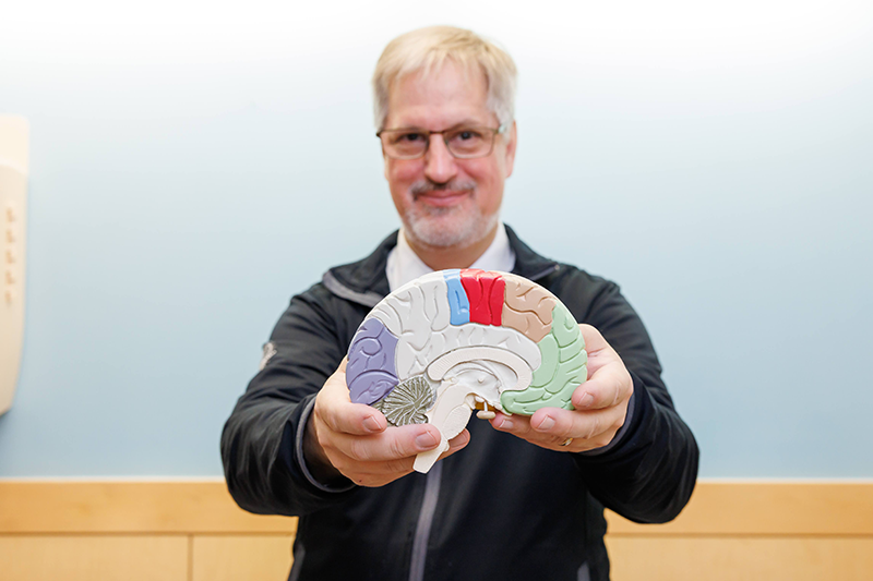
Browsing Category

Neurology & Neurosurgery

Inside Pediatrics, Neurology & Neurosurgery

Pioneering Surgery Spares Parents and Infants from Helmets

The Cleft and Craniofacial Center at Children’s of Alabama is one of the busiest in the country, with some of the most experienced physicians and support staff. From cleft palate to craniosynostosis (a condition in which the skull fuses too early) and complex tumor surgeries, the center draws patients from the entire Southeast region and beyond. It is a truly multidisciplinary group with neurologists, neurosurgeons, plastic surgeons, and a craniofacial pediatrician. 

It also offers state-of-the art therapies, including a new type of endoscopic surgery for craniosynostosis in infants as young as three months that is only performed in a few centers in the U.S. “The typical procedure is an endoscopic release of the craniosynostosis followed by post-operative helmet therapy,” said neurosurgeon James M. Johnston, M.D. “Helmet therapy works well, but kids have to wear it for 23 hours a day, and that can be a lot of work for families, especially when they live far from Birmingham,” he said. In addition, the Alabama Medicaid program, which covers most of these children, does not pay for the helmets, which can cost thousands of dollars (and children often need more than one). This puts tremendous financial strain on many families.  

So Dr. Johnston, joined by neurosurgeon Curtis J. Rozzelle, M.D., and plastic surgeons Rene’ P. Myers, M.D., and John Grant, M.D., brought spring-mediated cranioplasty, which was developed at Wake Forest University, to Children’s. It starts with the same endoscopic craniectomy used for children who would require helmets. Only in this procedure, the plastic surgeon steps in and inserts custom-made springs into the bony defect created by the surgery. The springs work to expand the skull over several months to correct the abnormal head shape and ensure appropriate cranial volume for brain growth. A few months later, the plastic surgeon removes the springs during a same-day surgery.  

“What’s nice is that there’s no need for a helmet,” Dr. Johnston said. Plus, studies show the procedure is just as safe and effective as cranioplasties requiring helmets.1 It’s also covered by all health insurance. “So, we’re able to do it for all children,” he said.  

A similar procedure using cranial distractors like those used to lengthen femurs is used for skull expansion, explained Dr. Grant. This technology is used in older children who need more intracranial volume but who are beyond the age at which the skull can form new bone to fill in surgically created soft spots. By “stretching” the bones of the skull more slowly, he said, the child’s body adjusts by making bone to fill in the growing gap. 

Regardless of the procedure used, early referrals are critical for these babies, said Dr. Rozzelle. “If we can see them by 2 months of age, that gives us plenty of time to get whatever preoperative assessments we need and get them on the schedule so that either the spring or endoscopic craniectomy with subsequent molding helmet is a viable option,” he said. Older babies cannot be treated endoscopically and require standard open surgery, which may lead to more blood loss and longer hospital stays.2 

Yet the craniofacial clinic still sometimes sees babies 6 months or older who never received a diagnosis or whose pediatrician didn’t refer them to Children’s. “That’s frustrating,” Dr. Rozzelle said. 

Nonetheless, said Dr. Myers, “Since we are comfortable with all of the techniques, we can tailor a plan to the individual child. No one is exactly the same.” 


1 Arko L, Swanson JW, Fierst TM, et al. Spring-mediated sagittal craniosynostosis treatment at the Children’s Hospital of Philadelphia: technical notes and literature review. Neurosurg Focus. 2015 May;38(5):E7

2 Hashim PW, Patel A, Yang JF, et al. The effects of whole-vault cranioplasty versus strip craniectomy on long-term neuropsychological outcomes in sagittal craniosynostosis. Plast Reconstr Surg 134:491–501, 2014.

Inside Pediatrics, Neurology & Neurosurgery

Advanced Imaging Enables Complex Surgeries for Epilepsy

If you’re going to conduct surgery on the brains of children with severe epilepsy, you better know what type they have, where they have it, and how it affects function.  

That’s where functional imaging comes in, including single-photon emission computerized tomography (SPECT), functional MRI (fMRI), positron emission tomography (PET), and magnetoencephalography (MEG). Most neurosurgical centers have one or two; but Children’s of Alabama has them all.  

“This is important,” said pediatric neurosurgeon Jeffrey P. Blount, M.D., “because there is never perfect alignment between the studies.” With multiple studies, however, comes greater certainty about the brain regions the disease impacts, which provides greater certainty about which parts to remove during surgery. Agreement between the scans is called “concordance,” and it is the central concept in epilepsy localization, said Dr. Blount.  

Most patients who require epilepsy surgery also require an invasive monitoring system prior to surgery, said neurosurgeon Curtis J. Rozzelle, M.D. In the past, he explained, that required an open cranial exposure to place electrodes on the surface of the brain and, sometimes, within the brain. 

But with newer techniques, particularly stereoelectroencephalography (SEEG), a minimally invasive surgical procedure used to precisely find the areas of the brain where seizures originate, surgeons can place an array of depth electrodes without performing a craniotomy. Instead, each electrode is placed robotically through a tiny hole drilled in the skull using a robotic stereotactic approach. “That relies very heavily on high-resolution scans,” Dr. Rozelle said, including fusing CT and MRI images, to put the electrodes in without damaging a critical part of the brain. 

“Mostly what we’re trying to avoid is hitting blood vessels with the depth electrodes while getting an array of electrodes that will cover the area of interest,” Dr. Rozelle said. The functional imaging studies are critical in establishing the target zones. Plus, since MEG and fMRI are based on magnetic field fluctuations, the MEG images can be mapped onto the MRI scan in three dimensions. The older technique, in which electrodes were placed on the surface of the brain, only provided a two-dimensional image. 

The child spends several days with the implanted electrodes to capture data about the seizures, which a neurologist then analyzes to identify the exact area of the brain that requires treatment. That surgery itself also relies heavily on high-resolution imaging. A laser ablation, for instance, is performed in the MRI scanner. A larger-volume surgery that requires open resection also relies on imaging because the surgical target looks the same as the normal brain. “To help us ensure that we hit the target, we can map the neurologist analysis into a navigation system that directs us to the right area,” Dr. Rozzelle said. “That ensures that we remove the tissue we need to take out and keep everything else intact.” 

Neurosurgeons at Children’s perform about 50 cranial epilepsy procedures a year, of which about 30 require the invasive monitoring. 

“We are very fortunate to work in a center where we have so much high-quality functional imaging available on a single campus,” said Dr. Blount. 

Inside Pediatrics, Neurology & Neurosurgery

Addressing Post-Traumatic Syndrome Disease from Hydrocephalus

Hydrocephalus-Doctor-Brain-Scans-Resized-V2

Children’s of Alabama neurosurgeon Brandon Rocque, M.D.

It’s not surprising that kids with brain tumors and their parents experience a significant amount of stress and psychological distress during the acute post-diagnosis period. It even has a name: pediatric medical traumatic stress. As Children’s of Alabama neurosurgeon Brandon Rocque, M.D., studied this phenomenon a few years ago, it occurred to him that it would almost certainly apply to children with hydrocephalus. 

“We know that just encountering doctors or the medical system can be traumatic for children,” said Dr. Rocque. “For children, just coming to the hospital can be traumatic enough to trigger post-traumatic stress disorder,” or PTSD. 

Numerous factors contribute to stress, particularly the perceived threat to the child’s life. “Even if there isn’t a threat, the child perceives it as such,” Dr. Rocque said. Add to that separation from their parents, uncertainty about the outcome, and the unpredictability of a serious medical condition. “That describes hydrocephalus extremely well,” he said, because these children are treated with shunts that could become blocked at any time requiring additional medical interventions.  

Symptoms of shunt failure can vary widely. Some children simply have a mild headache; other patients can become extremely sick and be in danger of death within a couple of hours. By age 10, “the average child [with hydrocephalus] has had at least two shunt replacements. This is always hanging over the families,” Dr. Rocque said, putting them and their children at high risk for PTSD. 

To test his hypothesis, Dr. Rocque introduced a screening survey into the hydrocephalus clinic to screen for PTSD as well as anxiety, depression, fatigue and resilience. “We found that, overall, the kids with hydrocephalus are doing pretty well. But the parents are not doing so well,” he said. About one in five parents met the diagnostic criteria for PTSD based on their symptoms. More than half attributed it to their child’s condition.  

So why aren’t the kids as affected? One reason, Dr. Rocque said, is that the children don’t know anything different. They’ve lived their entire lives with the condition and the shunts. “But for parents, there was always something new and the risk that something bad is going to happen to their child,” he said.  

Not all the kids surveyed were fine, however. “Some had issues with PTSD, and those were the ones coming to the hospital more. Those whose shunts weren’t behaving well,” Dr. Rocque said. “We need to be aware that these kids have a higher risk for PTSD.”  

They also found that the children and their patients tested exhibited very little resilience, which can help protect against PTSD.  

A survey conducted in conjunction with the Hydrocephalus Association confirmed their findings.  

Dr. Rocque and his team are now working with the association to develop a program to help reduce the risk of PTSD in patients and their families and with a psychologist who is also the mother of an adult with hydrocephalus to develop a tool to help build resilience in patients and their families.  

“This is the first time anyone has really focused on the psychological comorbidities of this condition,” Dr. Rocque said. “I think it has the potential to have a big effect in our population.” 

Inside Pediatrics, Neurology & Neurosurgery

Exploring the Brain from the Inside Out

Pediatric neurointerventional radiology is a small but growing specialty, one increasingly in use given the growing number of endovascular procedures performed in children with neurovascular conditions. “It’s a niche specialty,” says Jesse Jones, M.D., Children’s of Alabama Chief of Neurointervention. “A lot of doctors don’t know about it—let alone patients.”  

Dr. Jones is part of the hospital’s vascular anomalies team, one of the largest pediatric vascular anomalies programs in the Southeast and the only one in Alabama. He works with an interdisciplinary team of experts specializing in the diagnosis, treatment and ongoing care of all vascular anomalies and is part of the team’s monthly clinic. 

On the adult side, neurointerventional radiologists spend a lot of time removing blood clots from stroke patients. But stroke is rarer in children. The hospital’s neurosurgeons and neurologists more often call on Dr. Jones to evaluate congenital anomalies, including vein of Galen malformation or arteriovenous malformations (AVM), as well as inflammatory disorders like vasculitis or obliterative vasculopathy. “It’s when a child presents with dangerous or unusual neurovascular findings and the team is trying to characterize it and plan future treatment that I come in,” he said. 

Dr. Jones, who completed a residency and two fellowships, uses minimally invasive techniques to diagnose and treat numerous neurovascular conditions, including stroke and AVM, but also aneurysms, and lympho-vascular proliferations of the head and neckThe beauty of his approach is that it helps avoid open incisions, reducing the risk of complications and enabling kids to go home sooner. 

His interest in pediatric medicine started with his grandfather, who was a pediatrician. “I looked up to the work he did treating children,” Dr. Jones said. “Working with adults can get frustrating because many conditions they have could have been avoided with lifestyle changes. But in children, they bear no responsibility.” 

Dr. Jones also knew he wanted to do something with the brain. “I’m fascinated with how the brain works,” he said. “It’s a miraculous organ and even after all these years of study still a bit of an enigma.” Being involved in a neuroscience-related field and interacting with other specialists who study the brain is intellectually stimulating, he said. “It’s the best of both worlds: I get to use my hands as an interventional radiologist and work with the brain too.” 

And, of course, work with children. 

Inside Pediatrics, Neurology & Neurosurgery

Pediatric Neurosurgeon Launches Interactive Website Fostering Global Collaboration

Having partnered over the years with neurosurgeons in Vietnam and Ghana, James Johnston, Jr., M.D., a pediatric neurosurgeon at Children’s of Alabama and the University of Alabama at Birmingham (UAB), knew many in his field who craved this type of global collaboration but didn’t know where to start. That’s why he co-founded an interactive website designed to bring specialists and organizations together to improve the care of surgical patients worldwide.

Known as InterSurgeon (https://intersurgeon.org), the effort is the joint vision of Johnston and British pediatric neurosurgeon William Harkness, M.D., who focused on the stark fact that 80% of the world’s population lacks access to safe, timely and affordable surgical care. The pair, with support from multiple international organizations and Dean Vickers at the University of Alabama at Birmingham (UAB), raised funds to build InterSurgeon to help fill this void. It matches surgeons from disparate locations to not only collaborate, but form a supportive global community of like-minded professionals.

Initially launched with pediatric neurosurgeons in mind, InterSurgeon now also includes members from many other surgical specialties. The free service empowers surgeons, anesthesiologists, allied health professionals and equipment providers to partner on training, education and clinical care as well as share equipment and other resources.

“We’ve tried to create a stand-alone nexus for all players in global surgery to be able to join with others to better collaborate,” said Johnston, also an associate professor of pediatric neurosurgery at UAB.

“The World Health Organization passed a resolution in 2016 that put new emphasis on global surgery training as a major priority for global health,” he said. “We focus so much on infectious diseases, but what’s ended up happening is that the annual morbidity and mortality from surgery worldwide dwarfs all of that. It’s stunning.”

Key partnerships between InterSurgeon and other organizations over the past several years have driven opportunities for collaboration as well as access to surgical education with specialized technology. In addition to the United Nations Institute for Training and Research (UNITAR), partners include the G4 Alliance, which advocates for increased access to safe surgical care; and Ohana One, which has sent “smart glasses” loaded with augmented reality software from Birmingham-based HelpLightning to various sites around the world. This enabled mentor surgeons in developed healthcare systems to virtually interact in real time with mentees performing surgery for training purposes.

 With procedural competence integral to the specialty, surgery requires “a certain amount of infrastructure, and a lot of that has lagged worldwide,” Johnston noted. “But even in places with equipment, the quality of surgical training is not always as good as it could be.”

 With more than 600 members in 95 countries and growing, InterSurgeon aims to “shore up” that gap.            

“It’s a very difficult problem, and no one thing will solve it,” Johnston said. “But in surgery, it’s very important to connect experts and institutions with learners to bring them up to speed and improve the quality of the surgery they’re doing.”     

Inside Pediatrics, Neurology & Neurosurgery

Probing the Molecular Underpinnings of Undiagnosed Muscle Disorders

The Jerry Lewis fundraising telethons of yore educated many people about myopathies, the most famous of which – muscular dystrophy – highlights the often-progressive and disabling muscle weakness afflicting these children. But up to 10% of myopathy cases seen by Michael Lopez, M.D., Ph.D., a Children’s of Alabama pediatric neuromuscular physician-scientist, are undiagnosable, despite comprehensive evaluation.

This dilemma has driven Lopez, also an assistant professor of pediatric neurology at the University of Alabama at Birmingham (UAB), to collaborate with colleagues to use whole genome and RNA-Seq sequencing to potentially solve such cases. Lopez has enrolled 10 patients and their families into innovative clinical research aiming to reveal the molecular basis of undiagnosed myopathies in hopes of offering affected patients a prognosis and steering them toward effective treatments.

“In a small sliver of patients, I’m convinced they have a myopathy but am unable to refine their diagnosis and give clarity on what type,” Lopez explained. “In that group of kids, in which I’m pretty sure the cause is genetic and everything else is ruled out, we end up in this diagnostic odyssey.”

To tackle this problem, Lopez joined with UAB’s Liz Worthey, Ph.D., director of the Center for Genomic Data Sciences, and Matthew Alexander, Ph.D., an assistant professor of pediatric neurology. Using simple blood draws from parents and children and next-generation gene sequencing techniques, the trio hope to identify new genes responsible for muscle disorders or previously unreported variants of uncertain significance, dubbed VUS, in genes already known to cause myopathies. These mutations can be inserted into animal models to build evidence that a specific VUS triggers the condition.

“This allows us to get a snapshot of all the mutations possible in the genome, not excluding places that turn genes on and off,” Lopez said. “Data can help us support or refute a disease-causing mechanism for the VUS.”

Lopez is well aware the project won’t produce quick answers, and participating families also understand the findings may not benefit them personally.

“But it is possible to come back with a molecular diagnosis,” he said. “If we find something that’s already well-understood, that would offer them some treatment options if they’re already available.”

 Longer-term, Lopez hopes the research will point toward targeted treatments for these muscle disorders, some of which might be derived by repurposing old drugs in “off-the-shelf libraries.”            

“Treatment is just one goal, a second is resolving the diagnosis and solving the case,” he said. “That’s a huge burden relieved for both family and patient. And another piece is the science – to improve our understanding of how these diseases occur and how the muscle functions.”

Hematology and Oncology, Inside Pediatrics, Neurology & Neurosurgery

Children’s of Alabama Launches Second Groundbreaking Trial of Viral Treatment for Brain Tumors

Children’s of Alabama and the University of Alabama at Birmingham are leading studies using a genetically re-engineered herpes virus to treat pediatric high-grade gliomas.

“A uniformly dismal prognosis.” That’s how Children’s of Alabama neurosurgeon James M. Johnston, Jr., M.D., describes what children with recurrent malignant brain tumors face, with an average lifespan of six months given a lack of effective treatments.

Now Johnston, in collaboration with Greg Friedman, M.D., associate professor of pediatric oncology and director of Developmental Therapeutics at Children’s, is leading groundbreaking studies designed to shift that trajectory. The team recently completed a Phase 1 immunotherapy clinical trial of genetically re-engineered herpes virus G207 to treat pediatric high-grade gliomas. Their work builds on adult research on the viral treatment pioneered by James Markert, M.D., MPH, who chairs the Department of Neurosurgery at the University of Alabama at Birmingham (UAB), as well as Friedman’s laboratory work, which showed the virus was more effective against pediatric brain tumors than adult tumors.

In the past three years, 11 patients with high-grade gliomas have traveled to Children’s from throughout the country, Mexico and Canada to participate in the study. They receive special screening to pinpoint the tumor location, which is then biopsied. Johnston then places three to four catheters in the tumor. The next day, Friedman and his team infuse the genetically-modified virus into the brain through the catheters.

“We think the virus works by directly killing the tumor cells,” Johnston said, as well as activating the immune system to destroy any remaining cells. “Brain tumors have a way to hide from the immune system by making themselves immunologically ‘cold,’” he explained. The herpes virus turns a “cold” tumor into a “hot” tumor and generates the immune response. Indeed, months after the surgery tests show that immune cells have infiltrated the tumor and continue killing tumor cells.

The initial phase 1 trial in patients with high-grade gliomas was designed to demonstrate safety and wasn’t powered to show efficacy. Nonetheless, Johnston said, “our median survival was significantly longer than the historical six months,” with several children now long-term responders.

In late December 2019, the team received a three-year, $750,000 R01 grant from the U.S. Food and Drug Administration for a Phase 1 trial in malignant cerebellar brain tumors, which may be even more sensitive to the virotherapy than the gliomas. At the same time, they are submitting grants for a Phase 2 multicenter trial of the virus therapy for recurrent malignant supratentorial tumors.

Johnston stresses that the research is a team effort, involving basic scientists, oncologists, surgeons, nurses and intensivists. “It’s an ‘all-hands-on-deck’ kind of thing,” he said.

Inside Pediatrics, Neurology & Neurosurgery

Reducing Spasticity, Improving Quality of Life – One Child at a Time

Children’s of Alabama neurosurgeon Brandon Rocque, M.D., left, is part of a multidisciplinary team that offers surgical and medical therapies for children with cerebral palsy and severe spasticity.

Take orthopedic surgeons, neurosurgeons, rehabilitation medicine specialists, physical and occupational therapists, and specially trained nurses and what do you have? A multidisciplinary team that makes Children’s of Alabama one of the few pediatric medical centers in the U.S. to offer state-of-the-art surgical and medical therapies for children with cerebral palsy and severe spasticity, whether that be selective dorsal rhizotomy or intrathecal Baclofen pump therapies.

“We all work together to determine the best option for the child,” said Children’s neurosurgeon Brandon Rocque, M.D. But the medical professionals aren’t the only ones who play a role in the decision. “The most important factor is the family’s goals for the child,” Rocque said, which typically focus on some improvement in walking.

The two main options are rhizotomy, in which the surgeon exposes, stimulates and then severs nerve roots in the spinal cord, or a Baclofen pump, which continuously releases the muscle relaxer through the implanted pump.

“There are an infinite number of ways to stimulate the nerves (for rhizotomy) and make decisions about what to cut and not cut,” Rocque said. “Every program is unique.” To bring a more objective approach to the procedure, Children’s participates in the Cerebral Palsy Research Network (CPRN), a group of 25 medical centers committed to planning and executing high quality clinical research and quality improvement protocols.

“The idea of CPRN is to pull clinical data from the electronic medical record from children with cerebral palsy into a single data repository to understand it better,” Rocque said. That includes identifying children most likely to benefit from rhizotomy as well as the most appropriate surgical approach based on their condition.

For children who don’t require the extensive rhizotomy surgery, orthopedic surgery to release tight hamstrings or Achilles tendons might be used to reduce spasticity. And for those with more severe spasticity or dystonia, there’s the Baclofen pump.

More recently, the team has been performing palliative rhizotomy for children who would typically be considered for a Baclofen pump. While the pump remains an option, Rocque said, it is an implanted device, has to be refilled and carries a risk of infection and mechanical complications. Palliative rhizotomy, while not restoring the ability to walk, improves muscle tone in the legs. “Families are thrilled with how much looser the kids are, how much more comfortable and easier to take care of.”

The program sees about 100 children a year, most referred through rehabilitation medicine specialists, and hopes to expand from a bimonthly to a monthly clinic.

Inside Pediatrics, Neurology & Neurosurgery

Children’s of Alabama Clinicians Bring Expertise and Training to Vietnam

Global Surgery NEW_WEB

Children’s of Alabama physicians review a brain scan at a hospital in Vietnam. Neurosurgeons, neurologists and other medical staff travel to Vietnam at least once a year to provide lectures and hands-on training at hospitals in Hanoi and Ho Chi Minh City as part of Children’s of Alabama’s Global Surgery Program.

Surgical interventions to reduce the burden of drug-resistant epilepsy in children have become an integral part of the field in the past 20 years. In low- or middle-income countries like Vietnam, however, it typically remains a vision, not a reality. Vietnam, for instance, has just two adult and two pediatric neurosurgery training programs for a country of 95 million people, and just four pediatric neurosurgeons serving a population of more than 50 million in the northern part of the country.

Enter Children’s of Alabama’s Global Surgery Program, which is designed to form strong, collaborative relationships with large pediatric hospitals in low- and middle-income countries and provide subspecialty fellowship training at Children’s. The hospital’s relationship with Vietnam began in 2013, with an initial visit to neurosurgeons in Ho Chi Minh City. Since then, a team from Children’s, including pediatric neurosurgeon Brandon Rocque, M.D., MS, FAANS, Pediatric Epilepsy Surgery Director Pongkiat Kankirawatana, M.D., Clinical Neurophysiology and Pediatric Epilepsy Program Director Monisha Goyal, M.D., and Director of Neuromonitoring Trei King, R.EEG.T, C.N.I.M., among others, has traveled the nearly 9,000 miles to Vietnam at least once a year to provide lectures and hands-on training at hospitals in Hanoi and Ho Chi Minh City. Their efforts have helped create premier epilepsy programs that draw children from throughout southeast Asia.

“We started with the corpus callosotomy,” Rocque said, a procedure performed on children with generalized epilepsy prone to drop attacks. It involves splitting the main connection pathway between the two cerebral hemispheres to prevent the attacks. The Vietnamese team, led by a neurosurgeon who specialized in brain tumors, performed two such surgeries with the Children’s of Alabama neurosurgeons, then went on to complete 10 themselves over the next six months, all with good long-term results.

Epilepsy surgery is not possible without advanced EEG monitoring, which is where King came in. His job was to teach EEG technologists how to use an EEG in the operating room, including electrode placements, and the most appropriate test for the child’s condition. “We started with the basics and now we’re going much deeper,” he said.

“The people there are extremely hard working and very, very smart,” King said. “They just didn’t have the opportunity and education. The training with our staff allows them to see the entire gamut of what we do in the field and, hopefully, grow to do what we do.”

The Children’s of Alabama team usually spends a week in each city, giving lectures and assisting with the more complex surgeries. “We’re not trying to hammer out a bunch of cases,” Rocque stressed. “The model is not missionary surgery; it’s teaching and working on the patients they asked us to assist with in order to reach a goal of improving specific techniques for the neurologists and surgeons.”

In addition to building the team’s skills in epilepsy surgery, the team hopes to improve how pediatric neurosurgeons are trained in Vietnam, Rocque said. Currently, residents are trained in neurosurgery but don’t receive any formal pediatric training. “There is a really big opportunity to improve the way pediatric neurosurgery is taught in this region,” Rocque said. To help in that goal, Vietnamese physicians and EEG nurses now come to Alabama for several months for focused training on various procedures.

The partnership has continued to grow, with the Vietnamese doctors sending PowerPoint presentations on difficult cases for discussion at Children’s of Alabama’s weekly multidisciplinary meeting, during which the neurology team develops treatment plans. Now the team also discusses the Vietnamese patients. “When we started, we often had to ask for more information and make some changes to the treatment plan,” Rocque said. “But over the last year, their own recommendations have been spot on.”

The experience has been eye opening for the Children’s of Alabama clinicians. “I found a pediatric neurology and neurosurgery program making a valiant effort in diagnosing and treating one child after another with minimal resources,” said Goyal, who visited City Children’s Hospital in Ho Chi Minh City. The children slept on cots that spilled out into the adjacent roofed but open courtyards and hallways, she said. “There were no fans and in September temperatures in Ho Chi Minh were far from balmy.” She was also struck by the fact that the government does not allow the use of benzodiazepines, one of the most common medications used to control seizures in the U.S.

However, Goyal said, “the small number of clinicians do an admirable job with limited resources. They learn from textbooks, not from mentors, even though they have much fewer technical and pharmacological resources.”

“As a physician, this has been a very rewarding experience,” Goyal said. She, Rocque and King are planning another trip this spring to Ho Chi Minh City to continue helping the hospital develop of its own pediatric surgical epilepsy program.

King echoed Goyal’s comments. “It’s been one of the most rewarding things I’ve experienced in my career,” he said. “You see the big difference you make not only in the training, but in the impact on the patients receiving care that they would not get unless we’re there.”

“The patients and families are so appreciative,” King said. “I’ve never experienced that level of appreciation before. I think a lot of it has to do with realizing that without the partnership and collaboration we wouldn’t be able to do the surgeries.”

For instance, he recalls one family whose child was operated on returning to the hospital a year later with a picture of the child to express their thanks. “Those are the things they make this so rewarding,” he said. “When we see the difference it makes in the life of the patients and families and the joy that continues long after the surgery.”

Inside Pediatrics, Neurology & Neurosurgery

At Children’s of Alabama, Transitioning Spina Bifida Patients to Adult Care Begins at Age 13

Spina Bifida_WEB

Professional heavyweight boxer Deontay Wilder greets a patient while touring Children’s of Alabama’s Comprehensive Spina Bifida Program space in this 2015 photo. The multidisciplinary program treats about 500 patients a year.

Before the 1970s, about one in four infants born with spina bifida died. Today, thanks to advances in neurosurgery, genitourinary surgery, gastroenterology and physical medicine and rehabilitation, between 75 and 80 percent now survive into adulthood.[1]

But surviving doesn’t mean thriving. That’s what clinicians from Children’s of Alabama’s multidisciplinary Comprehensive Spina Bifida Program, which treats about 500 patients a year, learned when they joined specialists at the University of Alabama at Birmingham (UAB) to develop an adult spina bifida clinic. While there is a greater awareness in the pediatric community about the overall need for transitional programs, said Jeffrey P. Blount, M.D., FAANS, chief of pediatric neurosurgery at Children’s of Alabama, more is needed than simply transferring a patient’s medical records.

“Our patients were arriving unprepared not just for adult health care, but for adult life in general,” said Spina Bifida Program Coordinator Betsy Hopson, MSHA, who assists patients in the transition from pediatric to adult care.  “We’d watch them grow up and hear about their dreams but every year it seemed like the light inside them was becoming dimmer and their opportunities fading away.” By the time they arrived at the adult clinic, she said, many patients were depressed and their childhood dreams seemed to have disappeared.

“We thought, ‘We’ve got to do better with this next generation,’” Hopson said.

They, along with Children’s of Alabama neurosurgeon Brandon Rocque M.D., MS, FAANS, started with data, surveying 188 patients in the adult clinic about their perceptions of their own disability. More than half (56.4 percent) identified as permanently disabled, with the most important predictors of disability being poor bowel continence and low education.[2]

They also found that 70 percent of those surveyed were not working and, again, poor bowel continence and low education were determining factors, as well as diagnosis (open versus closed spinal dysraphism). Rocque emphasized that patients with daily bowel accidents were six times more likely to be unemployed, while those with weekly accidents were three times more likely to be unemployed.

Thus was born the Individualized Transitional Plan (ITP), which starts at age 13. The objective is to ready the child for adulthood by developing and achieving five goals, including bowel management and educational achievement. Goals also include transitional readiness (ie, do they know their medical history, can they make their own appointments and see the doctor without a parent present); meeting a parent-initiated goal (ie, showering without being told or learning how to cook); and a personal goal, such as learning to eat healthier. Hopson individualizes care to transform each goal into a measurable task and the results become a contract and framework for the next few years.

They also receive a transition binder that includes a career interest survey. “They complete it before they see me and then I help guide them,” Hopson said. She also ensures their career  goals are realistic. For instance, one patient said he wanted to be the Marines, which she knew wasn’t possible. Instead, she suggested a meeting with a recruiter to discuss civilian jobs in the military.

About 45 percent of patients fall into the age group for the ITP, which is part of the clinic’s Lifetime Care Model (LCM). The LCM begins prenatally and continues throughout the adult years.[3] Hopson developed the ITP, which has generated great interest from other clinics in North America who are struggling with the challenges of implementing a meaningful transition program.

Since starting the program, Blount said, clinicians from throughout the country have contacted the team at Children’s to learn how to start their own program. “There’s such a need for a supportive multidisciplinary clinic to support these patients,” he said.

“This model could be replicated for other children with chronic conditions,” Hopson said. “If we’re doing our jobs well, our children are going to be adults. So we have some obligation to get them there in a way that provides a good quality of life.”

Bowel Management Program

Children with spina bifida are born with neurogenic bladder and bowel issues. While urination can be managed through catheterization, with children as young as 3 years old learning to catheterize themselves, “bowel management is much trickier,” Hopson said. Gastrointestinal motility slows and constipation becomes a chronic issue. Unfortunately, she said, many families prefer constipation to regular movements because it’s easier to handle. But “once you set up that pattern it’s hard to break,” she said.  And bowel management, she said, “becomes the most important issue in whether they are able to get a job.”

Today, the children in the spina bifida clinic benefit from working with pediatric gastroenterologist Mitchell B. Cohen, M.D., and members of the Division of Physical Medicine and Rehabilitation. Cohen chairs the UAB Department of Pediatrics and is physician-in-chief at Children’s of Alabama. He became interested in the program after hearing Hopson speak. He developed a protocol beginning at age 4 to help families understand the importance of a bowel management program and try to end their reliance on constipation.

Nonetheless, it remains an “extraordinarily difficult problem,” Roque said. “There is no magic bullet.” Still, “with Cohen’s help we’re making inroads on this extraordinarily difficult problem that has profound impact that echoes through rest of a child’s care, including issues of self-image and self-worth.”


[1] Talamonti G, D’Aliberti G, Collice M. Myelomeningocele: Long-term neurosurgical treatment and follow-up in 202 patients. J Neurosurg 2007; 107: 368-86.

[2] Davis MC, Hopson BD, Blount J, et al. Predictors of permanent disability among adults with spinal dysraphism. J Neurosurg Spine. 2017 August ; 27(2): 169–177.

[3] Hopson B, Rocque BG, Joseph DB, et al. The development of a lifetime care model in comprehensive spina bifida care. J Ped Rehab Med. 2018; 11:323–334