Browsing Category

Inside Pediatrics

Nephrology

Leading-Edge Technology May Change Kidney Transplant Monitoring and Help Other Specialties

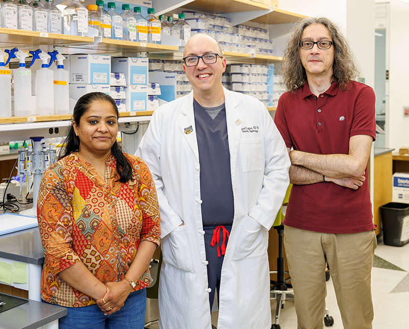
In the UAB Spatial Core Lab, researchers are using spatial transcriptomics to examine specific regions within tissue samples.

A breakthrough technology that allows doctors to study the precise regions of a kidney transplant that are involved in rejection could transform how doctors learn about kidney transplant biology, potentially leading to new diagnostic tests or treatments for children.

The technology, called spatial transcriptomics, is a leading-edge technique that lets researchers see which genes or proteins are active in very specific regions of tissue samples, such as those from a kidney biopsy. Unlike traditional methods that study tissue samples as a whole without regard for the location of important signals from the tissue, this technique allows researchers to examine custom-shaped regions of interest containing just a few cells in their natural environment.

Think of it as having a detailed map that shows not just what’s happening in a city, but precisely in which neighborhoods each activity occurs.

“There’s been decades now of data showing that gene expression patterns coming from a transplant are a little bit more sensitive for problems coming from a kidney transplant,” said Michael Seifert, M.D., director of the University of Alabama at Birmingham (UAB) Spatial Core and medical director of pediatric renal transplantation at Children’s of Alabama. “The problem is that we’ve never exactly known where those signals are coming from. Are they coming from cells in the kidney that we care about or are they coming from cells in the kidney that may not be as relevant?”

For instance, signals from immune system cells would be extremely relevant, he said, but could be distinct from those coming from the endothelial cells lining blood vessels.

With this technique, “we can look at a picture of a kidney biopsy on our instrument screen and take your mouse and draw a shape around it, and it will profile everything in that shape while ignoring everything else around it,” he said. It can even profile a certain cell type within the shape.

The Spatial Core team (from left): Pooja Nagaraj, MS, CCRP, Michael Seifert, M.D., Miguel Melendez-Ferro, Ph.D.

The technology itself isn’t destined for routine clinical use, Seifert said. “I can’t foresee a scenario where I would do a biopsy and then use spatial transcriptomics to make a diagnosis, because it’s a very labor-intensive, time-intensive and cost-intensive technique.”

Instead, he said, “my hope is that this will allow us to have a deeper understanding of the processes involved in transplants doing well but also transplants doing poorly. That will help us design better management programs, whether that’s using existing medicines in different ways or designing new medicines that can be more targeted and more effective than what we currently have available.”

Understanding exactly which parts of the kidney are affected by rejection also opens the door to personalized transplant care.

“Every cell in the kidney behaves differently depending on where it sits,” Seifert said. “This technology lets us uncover the heterogeneity—that is, the differences—within the tissue,” including if the problem lies in the blood vessels or the tubules or the parts of the kidney that generates urine. “I hope that’ll allow us to understand the signals that vary from person to person so we can really apply that more personalized technique.”

Thus, rather than treating all kidney transplant patients the same way, doctors could tailor anti-rejection treatments based on what is happening in an individual child’s kidney. This would, however, require advances in the spatial transcriptomics technology to make it faster and less expensive.

Spatial biology is not limited to the study of kidney transplant diseases. Seifert and his team in the UAB Spatial Core are working with specialists in other disciplines throughout Children’s and UAB, including ophthalmology, oncology and pulmonology. “We’re open to collaborating with any investigator with a good question that spatial biology can answer,” he said.

In fact, he sees spatial biology as an important technique for understanding all diseases in children. “I think what’s come out of this is an appreciation that the spatial context is incredibly important in so many of the diseases that we study.”

Orthopedics

How Dogs Are Transforming Pediatric Orthopedic Procedures

Dr. Michael Conklin and Shelby with a patient. (Photo courtesy of Judith Thomason)

What goes in must come out—including the metal pins used to hold bones together while fractures heal. “It probably takes 15 seconds to remove three pins,” Children’s of Alabama pediatric orthopedist Michael Conklin, M.D., said. “But, of course, kids are very scared about that.”

Enter Shelby. The 50-pound standard poodle is trained to sit on the examining table and cuddle with children while Conklin grasps the pins with a tool resembling a needle-nosed plier and pulls them out.

Basically, Shelby serves as a distraction, he says. “We tell the child to pet the dog and look toward the dog and not look at me on the other side of them, not worry about what I’m doing.”

Shelby remains calm no matter what, even with a screaming child. “She just sits there calmly and doesn’t do all the dog things that you and I know and love about our dogs,” Conklin said. “She’s trained to just be there for comfort.”

And it works. Well, for about two-thirds of patients. The rest “freak out no matter what,” Conklin said, but even then, Shelby has an effect. “It seems as if they return back to their baseline calm quicker after the procedure.”

Shelby also helps parents. “There’s a lot of value in the parents seeing that we’re trying to do our best for their child,” Conklin said. “Even though they know their child’s having to go through a procedure . . . it keeps us in good stead with them.”

Shelby’s brother, Foster, works with his sister at the Children’s South location as part of the Pups Unleashing Patient Smiles (PUPS) program, which is one of three branches of Children’s of Alabama’s animal-assisted program, PetsRX. Another branch involves longtime Children’s partner, Hand-in-Paw, which provides therapy dogs at Children’s of Alabama’s main hospital to provide comfort and distraction. The third is a hospital-based medical dog program which includes golden retrievers Wanda and Sydney to assist with scary or painful procedures. Meanwhile, suspected victims of child abuse—who are served by the Children’s Hospital Intervention and Prevention Services Center (CHIPS)—are assisted by dogs from the Help Empower Restore Overcome (HERO) Program with the Alabama Office of Prosecution Services.

The dogs aren’t just a cute addition. There is good science behind their use in the pediatric setting, with studies finding that animal-assisted therapy (AAT) can help children recover more quickly after surgery by improving mood and alertness, reducing perceived pain, and contributing to lower heart rates and blood pressure readings.[1],[2],[3] 

Animal-assisted therapy is also safe for the dogs, with studies showing no signs of stress or fatigue in therapy dogs when the programs are properly managed.[4] At Children’s, the dogs are overseen by a staff handler and rotated to ensure their well-being.

Soon, Shelby and Foster may be part of the scientific literature. Conklin and his team are conducting a randomized trial to evaluate the dogs’ effectiveness, comparing outcomes between patients who receive the therapy dog intervention and those who receive standard care. They are monitoring the child’s heart rate before, during and after the procedure to track how quickly they return to baseline and using a standardized anxiety scale that assesses facial expression, leg movement, activity, crying and consolability.

The goal is to, hopefully, show positive data that will pave the way for broader adoption of such programs.


[1] Calcaterra, V, Veggiotti, P, Palestrini, C, et al. Post-Operative Benefits of Animal-Assisted Therapy in Pediatric Surgery: A Randomised Study. PLoS ONE. 2015; 10.

[2] Braun C, Stangler T, Narveson J, Pettingell S. Animal-assisted therapy as a pain relief intervention for children. Complement Ther Clin Pract. 2009;15(2):105-109.

[3] López-Fernández, E., Palacios-Cuesta, A., Rodríguez-Martínez, A. et al. Implementation feasibility of animal-assisted therapy in a pediatric intensive care unit: effectiveness on reduction of pain, fear, and anxiety. Eur J Pediatr 183, 843–851 (2024). https://doi.org/10.1007/s00431-023-05284-7

[4] Palestrini C, Calcaterra V, Cannas S, et al.  Stress level evaluation in a dog during animal‐assisted therapy in pediatric surgery. Journal of Veterinary Behavior: Clinical Applications and Research. 2017; 17. https://doi.org/10.1016/j.jveb.2016.09.003.

Pulmonology

Bringing Asthma Care Closer to Home in Alabama’s Black Belt

Dr. Isabel L. Virella-Lowell with a patient.

Dallas, Marengo, Perry and Wilcox counties, part of the Black Belt (so named for its rich, dark soil), are four of the poorest counties in Alabama. They also have some of the highest rates of childhood asthma—nearly 12% compared to the state’s 8%—and are severely underserved when it comes to medical care. Two of the counties don’t even have a pediatrician.

Yet just 4% of the more than 5,000 patients seen at Children’s of Alabama’s Specialty Asthma Clinic hail from those areas. “We realized these kids weren’t getting to us for help,” pediatric pulmonologist Isabel L. Virella-Lowell, M.D., said. “But Medicaid data showed a high number of asthma-related claims from the area. So we knew there was a gap.”

One reason is distance, with families having to drive up to three hours to reach Birmingham. Thus, many children receive care only during asthma flare-ups at urgent care clinics or emergency rooms rather than ongoing, preventive treatment, said Children’s and University of Alabama at Birmingham (UAB) Pediatric Asthma Program Director Teresa G. Magruder, M.D. Without a primary care physician overseeing their child’s asthma, families find themselves caught in a cycle of crisis-driven care.

So instead of hoping kids will come to Birmingham, Virella-Lowell and Magruder are bringing their expertise to the Black Belt. Their mission: improve those dismal asthma statistics by engaging the community at a grass-roots level.

The initiative began when Children’s and UAB infectious disease specialist Claudette Poole, M.D., spent time in the area studying water sanitation and parasites. She kept hearing about an asthma crisis and recruited Virella-Lowell and Magruder.

The three applied for and received a Health Resources and Services Administration (HRSA) grant, which provides salary support for the core team and local community and health care partners. It also helps fund the virtual continuing medication education (CME) Project ECHO sessions, health fairs and supplies—such as spirometers and educational materials.

Magruder and Virella-Lowell stress the community-based approach of their efforts rather than having Children’s swoop in for just a few months. “We are really trying to improve the capacity of the community and their understanding of delivering asthma care in their own communities,” said Magruder. That means educating the front-line people caring for children, including school nurses, teachers, daycare workers and parents, while providing access to subspecialty care for the severe high-risk patients.

“If the community doesn’t buy in, if they are not engaged, if they’re not supportive, then there’s a limited amount of good we can do,” Lowell said.

“There’s some fatigue in these communities from programs that come and go,” Magruder added. “We know it takes time to build trust.”

The two doctors are also partnering with local physicians at Selma Pediatrics and Whitfield Regional Hospital in Demopolis. They hope to open a monthly clinic in 2026 in a space provided by Selma Pediatrics, so families don’t have to travel so far for specialty care.

They are also educating clinicians and others who see children with asthma through the aforementioned CME approach Project ECHO (which stands for Extension of Community Healthcare Outcomes), an interactive program that helps clinicians address their own cases. “It’s incredibly important that local physicians are comfortable managing asthma. And asthma care has changed a lot through the years,” said Lowell, who noted the recent release of new guidelines for diagnosing and treating the disease. The program, which is virtual, is available to any clinician throughout the state and beyond who’s interested in maintaining their expertise in pediatric asthma.

The Alabama State Asthma Coalition, a statewide group with a diverse mix of experts including environmental experts and respiratory educators, is also playing a role. The coalition helped train the first group of community health workers and hopes to continue that work.

Given that asthma affects one out of 10 children, it must be managed locally, Lowell said. “There’s no way that we will ever be able to manage all the really sick asthmatics here at Children’s. So it’s incredibly important that local pediatricians and family doctors are comfortable managing asthma.”

“Our goal is to raise the level of asthma care across the state,” Magruder said, “not just at Children’s, but everywhere kids need it.”

Endocrinology

Long-Term Effects of Gestational Diabetes on Kids

A new study from Children’s of Alabama shows the lasting impact of gestational diabetes on the child. (Stock photo)

New findings from a follow-up study at Children’s of Alabama and the University of Alabama at Birmingham (UAB) shed light on how a mother’s health during pregnancy may influence her child’s body weight well into adolescence—especially if that pregnancy was complicated by gestational diabetes. Led by pediatric endocrinology fellow Mary Margaret Barr, M.D., the new analysis builds on the foundational HAPi (Health After Pregnancy) study, conducted by  Paula Chandler-Laney, Ph.D., who directs UAB’s Ph.D. program in nutrition.

That original study assessed the health of 219 children ages 4 to 10. Mothers were divided into three groups based on her health during pregnancy: normal weight mothers without gestational diabetes (group 1); overweight or obese mothers without gestational diabetes (group 2); and overweight or obese mothers with gestational diabetes (group 3). Health-related data collected on the children included body mass index (BMI), waist-to-hip ratios, blood pressure and metabolic markers like glucose and cholesterol levels.

Barr’s research, which she presented at the Pediatric Endocrine Society annual meeting in May, went a step further. She reviewed electronic health records of 139 of the original study group to see how each group’s BMI Z-score—a metric that adjusts BMI for a child’s age and sex—changed as they entered adolescence.

As anticipated, children in group 3 (whose mothers had gestational diabetes and obesity) started off with higher BMI Z-scores that continued to trend upward through adolescence. “These were kids exposed to higher sugars while they were growing inside mom,” Barr said. Another key finding: Of all the children who had normal BMI Z-scores at the time of the original HAPi study (ages 4-10), those exposed to gestational diabetes (group 3) were significantly more likely to become overweight in adolescence.

Group 1—the control group—maintained healthy BMI levels over time, with only a slight rise in average BMI Z-score, which is often seen at adolescence.

The surprise came with group 2. These children, born to mothers with overweight or obesity but no gestational diabetes, initially had higher BMI Z-scores—even higher than group 3 at the study’s start. But over time, most of these children saw improvements in their BMI. “They started off big and then they got better,” Barr said. “Eventually, they ended up in the same range as the children born to normal weight mothers.”

This unexpected trend persisted even after adjusting for factors like maternal BMI, maternal education, household income and the number of children in the home. “These moms were of lower income, most of them below the poverty line, and had a lower education status,” she said. “You would have expected them to parallel group 3 and get worse over time. But they didn’t.”

The reason for the disconnect isn’t clear. “Nothing else stood out except for the child’s BMI during the HAPi study,” she said. “If you were heavier during the HAPi study, you were more likely to wind up heavier in adolescence. But it wasn’t a super strong correlation.”

Although none of the children developed diabetes during the follow-up period, Barr found a handful of prediabetes cases in groups 2 and 3.

The research provides a clue for pediatricians to intervene early in children with a high risk of obesity and/or diabetes. If the pediatrician knows the mother’s pregnancy weight and gestational diabetes history, they can be aware that the child may have a higher risk for obesity in the future. “So it’s probably more important to start earlier with healthy habits, a varied diet with less fried food, more vegetables, reasonable expectations of portion sizes, and exercise and movement,” Barr said.

While gestational diabetes and maternal obesity both increase a child’s risk for obesity, Barr’s findings suggest that gestational diabetes carries a more lasting impact than exposure to obesity alone. “We don’t fully understand the relationship between genetics, environment and exposures” on childhood obesity, she said. “But this data gives us another piece of the puzzle.” Her next step after publication is to expand the dataset to include maternal weight and metabolic health since the original study ended.

Gastroenterology

Children’s of Alabama Celiac Disease Clinic Growing to Meet Rising Demand

John Sands, M.D., leads the Celiac Disease Clinic at Children’s of Alabama.

Celiac disease is an autoimmune gastrointestinal disease that affects between 1% and 4% of the population. It is triggered by gluten, found in wheat, barley, rye and triticale (a mix of wheat and rye). Its incidence in children is increasing dramatically, with one study showing a 165% increase between 1994 and 2014, although recent studies suggest the incidence increase may have plateaued.[1],[2]

John Sands, M.D., who runs the Celiac Disease Clinic at Children’s of Alabama, is well aware of the increasing numbers, given the clinic’s own growth. In 2023, the clinic had 109 patient encounters. By 2024, that number had doubled to 221. Halfway through 2025, clinic staff had already had 157 patient encounters, on track for another record year. It’s gone from a monthly clinic to one that now meets three half days a month at different locations and times to make it more accessible to families.

The rising incidence in celiac disease is thought to be due to a combination of improved disease recognition and diagnostic testing, Sands said, as well as a true rise in the disorder itself. “Theories are that the increase is driven by environmental and lifestyle factors,” he said, including dietary changes and processed foods. Disruption of the intestinal barrier from viral infections and alterations in the gut microbiome (intestinal dysbiosis) are also being investigated as potential contributors in genetically susceptible individuals.

The core of celiac disease lies with a genetic mutation. But what’s puzzling is that while about 40% of the world’s population has the gene, only between 1% and 4% develop the disease, Sands said. “And probably half of those don’t realize that they’ve got celiac disease.” Most likely, he said environmental factors activate the gene.

More Than Stomach Problems

Many people misunderstand celiac disease, thinking it only causes digestive issues. “Celiac disease is not just belly pains,” Sands said. The condition can cause serious long-term health problems if left untreated, including bone loss, increased risk of heart disease, fertility issues and certain types of lymphomas.

The disease frequently appears alongside other autoimmune conditions. “There’s a triad of autoimmune diseases that tend to cluster together,” Sands said: Type 1 diabetes (T1DM), autoimmune thyroid disease and celiac disease.

In fact, many referrals to the clinic come from endocrinologists who treat children with T1DM and routinely screen for celiac disease. “It’s not uncommon for us to see those kids without any GI symptoms at all,” Sands said. Another clue the child may have celiac is poor growth. “It’s also not unusual for this to get picked up with kids who are shorter than we would predict them to be based on their parents’ height. You do the bloodwork for celiac disease and even though they have no GI symptoms, they have it.”

The Challenge of Living Gluten Free

The only treatment for celiac disease is strict, lifelong avoidance of gluten. This goes far beyond avoiding obvious sources like bread and pasta.

“Gluten is not just in food,” Sands said. “It’s all over the place. It’s in sunscreens. It’s in lip balm, it’s in shampoo, it’s in conditioners, it’s in Play-Doh.”

Following a gluten-free diet is not only challenging but expensive. He recalled one family with a child who had celiac disease that wanted the entire family to eat gluten free since the logistics of living gluten free would be easier for all than for one. But they couldn’t afford it,” he said. “A loaf of gluten-free bread can be six dollars.”

A Team Approach

The clinic’s core is its multidisciplinary approach. “One of the beauties of a celiac clinic is I have a registered dietitian,” Sands said, something that isn’t available to general pediatricians. The clinic also provides something else many doctors can’t offer: time.

“A general pediatrician is scheduling patients every eight to 10 minutes,” he said. “I’m setting up 40-minute slots and frequently spending longer than that with the patients and family.”

The clinic also participates in research efforts, including a collaborative study with Washington University examining immune changes in celiac disease progression.

Moving forward, Sands would like to provide more education about the disease for regional primary care clinicians, as well as patients and their families.

For now, he’s thrilled to be able to provide this specialized care, particularly since he follows his patients long term. “These are kids I get to know and see over and over again, and even watch go off to college.”


[1] Absah I, Patel B, Murray J, et al. Increasing Incidence and Altered Presentation in a Population-based Study of Pediatric Celiac Disease in North America. J Ped Gastro Nutr. 2017; 65 (432–437).

[2] VanNess GH, Ismail Y, Lee AT, King KS, Murray JA, Absah I. Are we beyond the peak of celiac disease incidence in Olmsted County, Minnesota, USA?. Gastroenterology and Functional Medicine. 2024 Dec 27;2.

Neurology & Neurosurgery

Zebrafish model shows potential XMEA treatments

A study in zebrafish of the ultra-rare disease XMEA could help researchers discover treatments. (Stock photo)

By Jeff Hansen, UAB

Can a small fish help identify possible treatments for an ultra-rare inherited disease found in an Alabama boy? The genetic disease is XMEA, which progressively weakens the muscles and can affect the liver and heart. As of March 2024, only 33 cases had ever been seen worldwide.

After the DNA sequence of the boy’s genome showed a mutation in the VMA21 gene, one of the known causes of XMEA, University of Alabama at Birmingham and Children’s of Alabama pediatric neurologist Michael Lopez, M.D., Ph.D., referred the family to the UAB Center for Precision Animal Modeling, or C-PAM.

At C-PAM and in collaboration with a Canadian group, research led by Matthew Alexander, Ph.D., UAB Department of Pediatrics, Division of Pediatric Neurology, and Jim Dowling, M.D., Ph.D., Hospital for Sick Children, Toronto, Ontario, created a preclinical model of XMEA in zebrafish by mutating the fish gene that is analogous to VMA21. While this small, striped fish is commonly found in home aquariums, zebrafish also are a valuable animal model for human disease due to fast growth, large clutch sizes and easy genetic manipulation. They also are transparent as larvae.

Matthew Alexander, Ph.D.

In a study published in EMBO Molecular Medicine, Alexander and Dowling now show that their mutant zebrafish have weakened muscles and other symptoms that mirror human XMEA disease. With this simple model, they were able to test 30 clinically tested drugs and identify two that significantly improved XMEA symptoms in the zebrafish. They now are studying the VMA21 mutation in a mammalian model, the mouse, to further push research toward a possible clinical treatment.

“We have established the first preclinical animal model of XMEA, and we have determined that this model faithfully recapitulates most features of the human disease,” Alexander said. “It thus is ideally suited for establishing disease pathomechanisms and identifying therapies.”

Researchers used CRISPR-Cas9, often called molecular scissors for DNA, to create two mutants: a frameshift mutation caused by a one-base pair deletion, and a premature stop codon created during deletion of 14 base pairs and insertion of 21. Both loss-of-function mutations reduced VMA21 protein levels.

Both mutants showed changes consistent with altered muscle structure and function, such as shorter body length and non-inflated swim bladders. They had reduced ability to swim away from a stimulus, and they spent less time swimming and traveled less distance compared to wildtype zebrafish.

The key cellular change in human XMEA is impairment of autophagy, the cell’s recycling system. Autophagy takes place in cell organelles called lysosomes, and these need to be acidic to activate proteases that degrade proteins for recycling into new proteins. Like human XMEA, the mutant fish lysosomes showed a failure to acidify, and the muscle cells had characteristic vacuoles — fluid-filled enclosed structures. Like human XMEA patients, the fish also showed liver and heart pathologies.

Unlike human XMEA, which can vary from mild to moderate symptoms as a progressive disease, the mutant fish showed severe reductions in life span, presumably due to a more complete loss of VMA function compared to human patients.

Since the fish had impaired autophagy and since there are no therapies for XMEA patients, the researchers tested 30 clinically tested autophagy inhibitory compounds from the Selleckchem drug library on the XMEA fish.

Screening of clutches for changed muscle birefringence, a change in the refraction of polarized light that indicates reduced muscle organization, the team identified nine compounds that both reduced abnormal birefringence and prolonged fish survival. Long-term testing of the nine for improvements in survival and swimming showed that edaravone and LY294002 had the greatest therapeutic effects.

“Excitingly, we found that several autophagy antagonists could ameliorate aspects of the VMA21 zebrafish phenotype, and two compounds in particular improved the phenotype across multiple domains of birefringence, motor function and survival,” Alexander said. “The fact that multiple autophagy modulators ameliorated aspects of the phenotype supports an important role for autophagy in the disease process and lends confidence to the validity and potential translatability of the findings to patients.”

Co-authors with Alexander and Dowling in the study “X-linked myopathy with excessive autophagy: characterization and therapy testing in a zebrafish model,” are Lily Huang, Rebecca Simonian and Lacramioara Fabian, Hospital for Sick Children; and Michael A. Lopez, Muthukumar Karuppasamy, Veronica M. Sanders and Katherine G. English, UAB Department of Pediatrics, Division of Pediatric Neurology.

At UAB, Pediatrics is a department in the Marnix E. Heersink School of Medicine.

XMEA stands for X-linked myopathy with excessive autophagy.

Neurology & Neurosurgery

Children’s of Alabama Offering New Gene Therapy for Patients With DMD

A new treatment offers the hope of longer, better life for DMD patients. (Stock photo)

In January 2025, Children’s of Alabama, for the first time, treated a patient with Duchenne muscular dystrophy (DMD) using a new gene therapy offered by only a few academic hospital facilities in the nation. The milestone followed a lengthy approval process and marked a new opportunity for patient success and scientific progress. Though not a cure, the treatment represents the hope of a longer life for these patients. For researchers, it will contribute to greater learning about the potential of this new treatment.

What is DMD?

While it is considered a rare disease, DMD is the most common form of muscular dystrophy, affecting one in every 5,000 males born in the United States. Patients experience progressive muscle degeneration, starting with proximal muscles and expanding to the limbs over time. They have trouble with many physical activities such as jumping, running and walking, and lose the ability to walk over time. The disease is fatal, and most patients don’t live past their late 20s. DMD has no cure; treatment focuses on extending the patient’s life by slowing down the disease’s progression.

According to the Muscular Dystrophy Association, symptoms of DMD can begin as early as ages 2-3 years, but in Alabama, where DMD is not yet part of newborn screening, many boys are not diagnosed until ages 4-6. That, says Children’s neuromuscular nurse practitioner Samantha Weaver, DNP, CRNP, is when patients begin to experience a steady decline. 

Treatment

Since the 1990s, physicians have prolonged the lives of patients with DMD using corticosteroids, whose anti-inflammatory properties can slow down the disease’s progression by about three years. Gene therapy, however, represents a new treatment aimed at restoring the function of the causative gene, DYSTROPHIN. The U.S. Food and Drug Administration originally approved it in 2023 for use in patients ages 4-5. In 2024, the agency extended that approval to all patients 4 years and older.

In this treatment, the transgene (a micro-DYSTROPHIN synthetic gene) is packaged within a viral capsid—a virus not intended to harm the patient that can hold the genetic material. In essence, physicians are “giving back the missing genetic information to the muscle tissue,” Children’s neurologist Michael Lopez, M.D., Ph.D., said. Unlike any other option, he noted, gene therapy treats the root cause of DMD.

“Now that we’re starting to get these really breakthrough therapies, they’re fulfilling on the promise that we all were searching for, which is that we could get closer to making this disease really better,” he said.

For the patient, improvements don’t happen overnight. That’s not how gene therapy works, Lopez says. “What we hope is that over many years, we’ll see a slow progression of the disease that is beyond what we would get with just treatment with corticosteroids alone,” he explained. “And I think that added benefit is something that’s going to be more of a long-term improvement.”

So, what can the parents of each patient treated with gene therapy hope to see? Ideally, in the short-term, their child will be more active. “I hope that our children can have more of a shot at more play and more jumping and more climbing and all of those things in the future,” said Erin McLeod, M.D., a pediatric neuromuscular neurologist at Children’s. In the long-term, the hope is that they’ll have a longer life.

Evidence supports the treatment’s efficacy. The clinical trials show that gene therapy is being delivered to patient’s muscles, and while the motor assessments haven’t shown clear evidence of clinically observable benefits, the data has trended toward improvement. Lopez says in other, more-recent studies, treated patients are starting to show improvements compared to those not receiving gene therapy. “The MRIs of the muscles themselves look a little bit healthier in some of these patients,” he said. “There’s less evidence of disease in that.”

From left: Samantha Weaver, DNP, CRNP; Erin McLeod, M.D.; Michael Lopez, M.D., Ph.D.

Finding the Right Fit

Gene therapy, however, is not the right fit for every patient. To determine candidates, Children’s looks at age, underlying disease and disease progression, Weaver said. They also consider the patient’s overall health and risk for infectious diseases. “It’s an extensive process,” she explained.

With all gene therapies, safety must be prioritized. The treatment can produce a significant immune response that can even prove life-threatening to patients with more advanced stages of the disease. Liver injury is also a major concern. Thus, Weaver says the team must ensure the patient has no antibodies that will reject the virus. “These are important steps to make sure the patient will have the best outcome,” she said.

Because of these considerations, only a small percentage of patients are ideal for the treatment.

Why Offer it at Children’s?

In Alabama, Children’s is the only hospital that offers gene therapy for patients with DMD. Making it available made sense—the hospital already treats spinal muscular atrophy (SMA) patients with gene therapy. Brad Troxler, M.D., and Shelley Coskery, CRNP, led the way on that, Lopez said, and “built in a lot of the infrastructure that we needed to be able to start doing gene therapies.”

“That really has put us out in front of the field with the experience to deliver these high-cost and novel leading-edge treatments,” he added.

Challenges

Cost was one of many challenges for the team as they sought approval to implement this multimillion-dollar therapy. To that end, they involved hospital administration and a pharmacoeconomics committee in the process. But, as Weaver pointed out, the process involved many more steps including determining who would write a protocol to ensure patient safety. They also had to build a larger team, which ultimately included hepatologists, pulmonologists, cardiologists, physical therapists and social workers.

Obstacles persist, even as Children’s offers the treatment. “A high cost remains a big challenge,” Lopez said. “And so we’ve been fortunate to be able to provide these treatments because we’ve gotten support from the insurers, so far.” But not every insurer is the same, he noted, and some may be slow to cover or even decline to cover the treatment.

What the Future Holds

So far, Children’s has dosed only one patient—out of roughly 100 that it follows—with the new gene therapy. While few will be candidates for the treatment, the team hopes to dose more in the future, as long as “the risk is appropriate and the benefit is continuing to be demonstrated,” Lopez said. The team also expects more advancements, which may make it possible for others—especially those with more severe cases—to receive the treatment.

“This first approved treatment for Duchenne that is a gene therapy is just the beginning, and there are going to be more down the road,” Lopez added. “There are certainly some that are in clinical trials now. So I think we’re right to be optimistic in that we’re starting to really push the treatment of Duchenne in a way that’s going to give us lots of options that weren’t there before.”

And as Children’s continues to offer the treatment, they’ll contribute to the scientific community’s information on its effectiveness, which means the team is paving the way toward greater success for the broader population of patients with DMD.

“I think everyone who is familiar with it at this point knows it is not a cure. But it is supposed to significantly slow the disease, and we are still gathering and gaining more data to that,” Weaver said. “So we’re very excited to be part of that process.”

Urology

A new protocol for kidney tests in spina bifida patients

A new study shows that ultrasound is not enough to monitor kidney health in children with spina bifida. (Stock photo)

For decades, doctors have relied heavily on ultrasound scans to monitor kidney health in children with spina bifida, the most common permanently disabling birth defect in the U.S. People with spina bifida tend to develop end-stage renal disease up to 20 years earlier than the general population, so keeping a close watch on kidney health from a young age is important, says Children’s of Alabama pediatric urologist Stacy Tanaka, M.D. “Then if there’s a concern, it can be acted upon early and not ignored.”

Current guidelines from the Spina Bifida Association (SBA) recommend annual screening with ultrasound to look for hydronephrosis—a condition in which the urine backs up into one or more kidneys—as a sign of kidney function, and blood tests like serum creatinine, to measure overall kidney health. But with kids, Tanaka says, “the practice pattern was that a lot of people were only doing renal ultrasound.”

Now a new study from Tanaka and her Children’s colleague David Joseph, M.D., as well as other kidney experts from around the country, shows that ultrasound alone is not enough to assess kidney health. “We basically use ultrasonography as a reflection of renal function,” Joseph said, but few, if any, studies assessed its accuracy in determining renal function.

Stacy Tanaka, M.D.

The study’s genesis came during a multidisciplinary meeting in 2003 of specialists who treat children with spina bifida. “The bottom line from all disciplines at that time was that nobody was really treating this population with evidence-based care,” Joseph said.

To change that, the Centers for Disease Control and Prevention and the SBA established the National Spinal Bifida Patient Registry (NSBPR), to which 20 spina bifida clinics submit data to help develop evidence-based care. In addition, nine clinics established a urologic protocol to manage and preserve initial renal function in young children with spina bifida (UMPIRE). The NSBPR and UMPIRE provided the data set Joseph and Tanaka used to determine the effectiveness of renal ultrasound vs. blood test to assess renal function.

The two registries included data on 2,500 children ages 1-18 with myelomeningocele, the most severe form of spina bifida. All had had an ultrasound and blood test within six months to determine estimated glomerular filtration rate (eGFR), a marker of kidney health.

The results were striking: ultrasound-based detection of hydronephrosis had only about a 25% sensitivity for identifying children with signs of chronic kidney disease in the UMPIRE study and 24% in the NSBPR cohort. That means kidney damage in three out of four children was going undetected. The poor sensitivity held even when researchers looked only at severe hydronephrosis, which had an even worse sensitivity rate–just 6% to 11%. “The renal ultrasound by itself wasn’t all that good,” Joseph said, “but that didn’t surprise us.”

David Joseph, M.D.

The findings challenge current practice and suggest that blood tests measuring kidney function should be routinely performed alongside ultrasound, not just when ultrasound results look concerning, as some clinicians practice. The team at Children’s prefers testing for cystatin C rather than creatinine because of the test’s improved and more accurate ability to obtain an eGFR.

One reason clinicians may eschew blood tests is that it involves needles, Tanaka said, which be traumatizing for children. Ultrasound, on the other hand, is noninvasive, easily available, and can be performed by technicians.

“The ultrasound is very helpful and important,” Joseph said, “but you need to recognize that it may not be telling you about renal function or injury to the kidney.” The findings have already changed practice at Children’s, where all kids with spina bifida now receive both tests during kidney health screening.

Ideally, the next study would randomize kids to either double testing or ultrasound alone, but that requires significant funding, particularly since the children would need to be followed for years.

This study was conducted with very little financial support, Tanaka said. “It represents a labor of love for everyone at all nine UMPRIE centers who have been involved in this project,” Joseph added.

Nephrology

Reducing hypertension numbers in children—and adults

The Children’s of Alabama Hypertension Clinic aims to help children in the short-term and long-term. (Stock photo)

Successfully stemming rising rates of cardiovascular disease in adults needs to start in childhood. But with mounting numbers of children and adolescents developing high blood pressure—a trend driven largely by skyrocketing obesity rates—this objective is getting harder to achieve. Enter the Pediatric Hypertension Program at Children’s of Alabama, which, with its steady growth, seeks to break the cycle.  

The Hypertension Clinic, which operates three half-days each week, now sees about 45 patients weekly, a 10-fold increase from 14 years ago, says Daniel Feig, M.D., Ph.D., director of the Division of Pediatric Nephrology at Children’s, who was recruited in 2011 to oversee the clinic’s development and expansion.

High blood pressure—defined in adults and children 13 years and older as a reading of 130/80 mm Hg or higher—is relatively unusual in healthy young patients, affecting 2-3% of typical children and adolescents. (For younger children, the definition of hypertension is a statistical one, based on greater than 95th percentile for age, sex and height.) But children with obesity—who account for nearly 20% of all Americans under 18—have a 20-30% rate of hypertension, says Feig, also the Margaret Porter Professor of Pediatrics at University of Alabama at Birmingham (UAB).

Daniel Feig, M.D.

“There’s a fairly large number of kids with high blood pressure, and one of the major concerns is how we can impact the long-term epidemic that results in cardiovascular disease in adults,” he said. “Controlling hypertension in adults hasn’t gone very well—only about half of those diagnosed have even remotely effective control. This impacts their cardiovascular disease and stroke risk.”

“The only way we can get this under control is by prevention,” Feig added. “If we can make an impact in children and adolescents, we can push back cardiovascular disease in adults.”

Drawing patients from across Alabama as well as some from eastern Mississippi and western Georgia, the Children’s Hypertension Program provides ongoing care for about 2,200 children. But this “catchment area” is likely home to about 70,000 young people with high blood pressure who are undiagnosed. Part of the problem is that many pediatricians aren’t comfortable diagnosing or treating the condition, Feig explains.

When patients come to Children’s, they’re often set up with ambulatory blood pressure monitoring equipment they wear for 24 to 48 hours to measure blood pressure a few times each hour while doing normal activities. The technology enables Children’s clinicians to tease out who actually has hypertension and not blood pressure spikes resulting from factors such as exertion, nervousness or pain.

Once diagnosed, Feig and pediatric nurse practitioner Jessica Edmondson collaborate with dietitians, pharmacists, social workers and others at Children’s to ensure patients benefit from a multidisciplinary approach to treatment. Ultimately, they’re trying to prevent both short- and long-term health implications resulting from hypertension, including heart thickening, retinal damage and even impairments in cognitive function.

It’s a daunting task, Feig acknowledges. “Anything we can do to reduce the numbers right now has a domino effect that reduces long-term target organ damage and long-term cardiovascular risk,” he said. “We’re not at a point where we can reverse the trajectory in 70,000 undiagnosed kids in Alabama, but we can positively impact a good number of kids, improve their health and quality of life, and gain the opportunity to gradually access more and more of them.”

Neonatology

Extended CPAP Shows Promise in Preemies

Research at Children’s and UAB shows that keeping premature infants on CPAP longer may improve lung growth.

Research at Children’s of Alabama and the University of Alabama at Birmingham suggests that keeping premature babies on nasal continuous positive airway pressure (CPAP) longer than currently practiced could significantly improve respiratory outcomes, potentially changing how neonatologists approach respiratory care for the smallest patients.

Early use of CPAP is standard for preterm infants unable to breathe on their own. It helps avoid invasive ventilation and minimizes the risk of lung injury while also increasing lung volume, which could stimulate lung growth and development.

Zaki Yazdi, M.D.

Children’s neonatologist Zaki Yazdi, M.D., conducted a pilot study as part of his fellowship to see whether extending CPAP beyond traditional stopping points benefited premature infants. Yazdi’s study, published in Archives of Disease in Childhood: Fetal & Neonatal, showed that continuing CPAP reduced episodes of drops in heart rate and oxygen levels in preterm infants. These positive findings align with groundbreaking research published this year in the American Journal of Critical Care Medicine, suggesting that extending CPAP promotes lung growth in babies born prematurely.

“We know CPAP helps premature babies with respiratory distress syndrome and apnea of prematurity,” Yazdi said. “The question we were trying to answer was: When is the best time to stop CPAP? We hypothesized that if you were to continue CPAP for a 24-hour period instead of going down to nasal cannula, you would have fewer drops in your oxygen level.”

Yazdi and the Children’s neonatology team, including neonatologist Colm P. Travers, M.D., randomized 36 infants born before 34 weeks gestation to either stop CPAP when they met Children’s traditional criteria (minimal oxygen support and few apnea episodes) or continue for an additional 24 hours. The primary outcomes were oxygen levels and other vital sign changes.

Colm Travers, M.D.

Babies who remained on CPAP an additional 24 hours experienced significantly fewer episodes of intermittent hypoxemia—defined as oxygen saturation below 85% for 10 seconds or longer—compared to those transitioned to low-flow nasal cannula. The CPAP group also had fewer heart rate drops and spent less time with low oxygen saturations.

“Even though all the markers we traditionally look at say this baby should be ready to come off CPAP, perhaps there are some more subtle things that we wouldn’t normally pick up on that suggest staying on CPAP could be helpful,” Yazdi said.

Extended CPAP isn’t without risks. Prolonged treatment can delay oral feeding, since many hospitals avoid feeding babies while on CPAP. There’s also risk of nasal breakdown from the CPAP mask interface, increased costs, and potential complications like feeding intolerance from swallowing air. However, Yazdi’s study found no negative effects during the 24-hour extension period.

The team has now received funding from the National Institute of Child Health and Human Development’s Neonatal Research Network to perform a much larger study examining extended CPAP’s effects on lung development. The multicenter, randomized clinical trial—led by UAB—will involve approximately 860 babies. Unlike Yazdi’s 24-hour study, neonates randomized to the longer CPAP arm will remain on the support for at least two weeks or until they are 34 weeks post-menstrual age. The children will then be followed for two years to assess lung function development and long-term respiratory outcomes, making it the largest controlled trial of extended CPAP to date. 

Already, Yazdi said, he and other neonatologists have noticed “a little bit of creep” toward keeping babies on CPAP longer at Children’s and other institutions.

“I don’t think we’re ready to say that this is definitely the best way to go yet,” Travers said. “But preliminary data that’s very promising suggests we need to do this larger trial to see if there’s any long-term benefit.”

“This could redefine what the standard of care could be,” Yazdi said.