Browsing Tag

neurosurgery

Neurology & Neurosurgery

McRae Addressing PTSD in Kids With Hydrocephalus

Elizabeth McRae, Ph.D., is a psychologist embedded within the Children’s of Alabama Neurosurgery team.

The young child was beyond terrified of the hospital. Born with hydrocephalus, he’d had numerous surgeries, and his anxiety was so high that just getting him to the car for doctors’ appointments was a struggle. It could take an hour to get from the parking garage to the hospital entrance given his tantrums and refusal to walk. The behavior continued at home every time someone opened the front door. “The parents really couldn’t live their life because it was so intense,” said Elizabeth McRae, Ph.D., a pediatric clinical child psychologist at Children’s of Alabama.

Here was a clear case of post-traumatic stress disorder (PTSD) related to the boy’s illness. Resolving it is exactly what McRae, who joined the neurosurgery team in January 2024, was hired to do.

For years, neurosurgeons and families caring for children with hydrocephalus understood the physical stakes: shunts that could fail without warning, repeated surgeries, emergency trips to the hospital. What was less visible—and often unaddressed—was the psychological toll of living in constant vigilance both for the patient and the family.

Earlier work at Children’s helped bring that reality into focus, documenting high rates of medical post-traumatic stress among families coping with hydrocephalus. But identifying the problem was only the beginning.

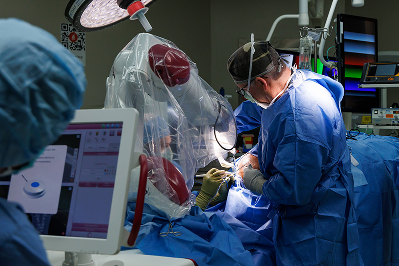
“Based on the results of that previous survey, we brought Dr. McRae on board and embedded her in our neurosurgery practice to provide psychological support for PTSD from screening and diagnosis through interventions to getting people plugged in to community resources,” pediatric neurosurgeon Brandon Rocque, M.D., said.

What’s emerged is an integrated, trauma-informed model of care that treats psychological health as part of standard neurosurgical practice.

From Measuring Stress to Building Resilience

Families of children with hydrocephalus face a unique kind of uncertainty, McRae said. Even when a child is medically stable, the possibility of sudden deterioration and a need for a new shunt never disappears. That’s why resilience, which she defines as strengthening the ability of families and patients to view difficulties as challenges rather than barriers, is so important.

Whether she’s meeting a family for the first time at diagnosis or after a child’s 10th surgery, she starts from the same place: helping them identify strengths they already have that can enable them to cope.

McRae also emphasizes connection. “One of the key predictors of potential traumatic stress is feeling like we’ve lost power and feeling isolated,” she said. “So if, right off the bat, we can empower them and encourage a connection, to me, those are two of the best things we can do up front.”

“There’s also a big piece of how do we prevent the trauma?” she continued. One approach, she said, is “taking a trauma-informed approach to our service so we mitigate the risk on the front end.” That includes explaining what’s going to happen to children; giving them options and a sense of control whenever possible; creating a sense of structure and predictability in the hospital setting as much as possible; and relying on other services such as Child Life to help children cope and adjust through play. 

McRae works closely with the surgeons, nurses and residents in both clinic and hospital settings, participating in the morning clinical discussions. Nurses refer families they see struggling, and surgeons seek her input about how to provide trauma-informed care in communication and interactions with patients.

The clinical model emphasizes brief, targeted interventions—an intentional choice in a population already burdened by multiple appointments. “I’m doing them a disservice if I can’t do something fairly efficiently,” McRae said.

These strategies were helpful with the aforementioned young child. McRae worked with him and his mother using developmentally appropriate coping strategies such as play-based breathing exercises, predictable reassurance and gradual exposure. She had him pretend to be a snake and breathe in slowly and deeply like a snake to quell his anxiety. A scavenger hunt throughout the hospital helped provide distraction so he could become comfortable in the medical setting. And Rocque met with him dressed in his blue scrubs for a meet-and-greet, no medicine involved, since the child was usually so frightened by anyone in blue scrubs. McRae also involved the boy’s mother in the interventions, providing a greater sense of control over the situation.

The result? The walk from the car to the hospital takes just a few minutes. The tantrums in the clinic are over. His parents have space to breathe. All this was achieved over the course of just six, one-hour sessions.

Research is also a big part of the program, McRae said. To that end, she and the team are collecting data on how the model functions, including who benefits most, how referrals happen, what interventions are feasible, and whether the approach is sustainable.

“This is the first time anybody has tried to integrate psychology into pediatric neurosurgery like this,” Rocque said. “So there are so many questions that we need to answer.”

That includes developing screening tools to identify which families need support most urgently and tracking service metrics to ensure the model can be replicated.

“We really want to show that this works,” Rocque said.

Early signs suggest it is. The model has already been adopted in other specialty clinics, including tuberous sclerosis.

“Ideally,” Rocque said, “I would love for this to become the standard of care in pediatric neurosurgery.”

Neurology & Neurosurgery

An endoscopic approach for skull base conditions

Dr. Jessica Grayson (left) and Dr. James Johnston perform an endoscopic procedure on a patient at Children’s of Alabama.

A growing number of children with complex skull base conditions can now be treated with minimally invasive surgery at Children’s of Alabama thanks to a collaboration between pediatric neurosurgeon James M. Johnston, M.D. and otolaryngologist Jessica Grayson, M.D. Together, they lead an integrated pediatric skull base surgery program that offers endoscopic procedures for conditions such as skull base tumors, traumatic injuries, complex pituitary lesions, and congenital abnormalities such as encephaloceles—in which brain tissue protrudes through an opening in the skull.

“Endoscopic approaches have been part of pediatric neurosurgery here for years for things like hydrocephalus or intraventricular tumors,” said Johnston, director of the Division of Pediatric Neurosurgery at Children’s and the University of Alabama at Birmingham (UAB). “What’s new and exciting is how we’ve expanded endonasal skull base surgery through this collaboration.”

The procedure involves threading a tiny camera and instruments through the patient’s nasal passages to reach the brain. “That means smaller incisions, less blood loss and a much shorter recovery time,” said Grayson—one of the few clinicians in the country who is fellowship trained in rhinology and skull base surgery for both adults and children, with extensive expertise in endoscopic endonasal surgery.

After tumor removal, Grayson works to patch any small holes created between the brain and the nose. This is one of the most critical aspects after the removal—if the small holes aren’t properly sealed, cerebral spinal fluid could leak out into the nose, leading to a high risk of infection. Grayson typically uses a nasoseptal flap to close any openings. She peels a small piece of the mucosa covering the nasal septum while maintaining its blood supply, then flips it over to cover any holes created during surgery.

The program is multidisciplinary, involving ENT, neurosurgery and occasionally plastic surgery. The team-based model also allows for comprehensive case review and planning. “We often consult with our adult colleagues at UAB when a case is really complex,” Johnston said. “It’s like having a built-in tumor board.”

Offering this type of approach for children is another way the program is unique—this method typically has been reserved for adults. And “the technical aspects are different from adult cases,” given their smaller anatomy and less-developed sinuses, Johnston noted. “But with collaboration, it’s absolutely feasible. We’ve even done this in infants as young as a few months old.”

The first collaboration—a case of congenital encephalocele in which the protruding tissue was initially mistaken for adenoid tissue—highlighted the potential of combining expertise. “That was the moment we realized we could safely and effectively treat these cases together using a minimally invasive endoscopic approach,” Grayson said.

Nationally, this type of program is rare. “In many places, kids are sent to adult hospitals for these procedures,” Grayson said. “Here, they can stay in a pediatric environment with pediatric anesthesiologists, nurses and postoperative care, which is crucial for safety and comfort.”

Last year, the team did about 40 cases, and the number of referrals is growing as more clinicians become aware of what’s possible. “We’re seeing more cases from outside hospitals,” Johnston said. “And we’re better at recognizing which patients are good candidates.”

Neurology & Neurosurgery

New MEG at UAB to enhance neuroimaging possibilities 

A new magnetoencephalography could improve treatment of multiple brain diseases at UAB and Children’s. (Photo by Andrea Mabry)

By Katherine Gaither, UAB

The complexity of the human brain has long been an enigma that neuroscientists have sought to untangle. Now, new technology at UAB will act as a critical tool to help researchers and clinicians interpret the brain in unprecedented ways.

UAB has recently invested in a new MEG, which stands for magnetoencephalography. It is used on pediatric and adult patients, so it benefits patients at both UAB and Children’s of Alabama. Put simply, MEG technology measures the magnetic fields that come from the brain’s nerve cells in an effort to analyze their function—and does so at millisecond intervals.

These implications are significant not only for localizing abnormalities in the brain in patients with diseases like epilepsy but also for studying how the brain performs normal functions like speaking, hearing, and seeing.

“It’s not invasive,” said Ismail Mohamed, M.D., professor in the UAB Division of Pediatric Neurology, Department of Pediatrics. “You don’t have to put electrodes in the brain, and it has no risks. You can potentially measure brain activity across multiple sessions. You can potentially measure them across a lifetime span. You can use it to learn things about how our brain functions.”

Measuring the brain’s magnetic fields

UAB was among the first medical centers in the country to obtain a MEG, having done so originally in 2001; however, evolving technology has created a need for replacing the old technology with a new one. The new machine was installed in September 2024.

Many are familiar with MRI as a form of imaging to interpret brain activity; however, having a MEG is not as common. UAB is one of fewer than 30 clinical centers in the nation that houses this technology.

“MRI looks at structure, but MEG primarily looks at the brain waves itself,” Mohamed explained.

The machine operates in a sealed room with a thick door, which eliminates outside magnetic noise. Patients lie or sit still during the scan, which takes precise magnetic field measurements of brain activity.

“The experience is not much different from laying inside an MRI scanner; however, the technology is quite different, and the way we measure is quite different,” Mohamed said. “It’s a passive measurement, which means that even if you’re pregnant, for example, you still can get a MEG scan. There are no risks.”

Compared to MRI and other brain scans like PET, and SPECT, the MEG gives you unique information about the brain as it tracks the activity of the nerve cells. EEG scans are similar, but the MEG has a heightened ability to localize this activity.

“A traditional EEG uses 25 electrodes. The MEG has 306 sensors,” Mohamed said. “So that coverage of the brain is bigger, it enhances the potential to produce more accurate information.”

According to Benjamin Cox, M.D., assistant professor in the UAB Department of Neurology, the difference is also electric vs. magnetic.

“The electrical fields that EEGs are recording are very much attenuated by the skull and all the intervening tissues,” Cox said. “The magnetic fields are not. So, we get a lot more precise localization with the MEG.”

Clinical implications

One significant implementation of the MEG is for use in epilepsy surgery to determine where in the brain seizures originate. Surgeons can use the results of a MEG scan to plan epilepsy surgeries.

“When we’re doing epilepsy surgery and trying to figure out if patients are a surgery candidate, we need to know exactly where the seizures are coming from as precisely as possible, and many times we end up putting electrodes in the brain to sample that activity directly,” Cox said. “So having studies like MEG, where we can have a precise idea of where to put those electrodes, is very helpful.”

Kristen Riley, M.D., professor in the UAB Department of Neurosurgery, notes that “MEG studies help us as surgeons to localize seizure onset zones, directing us to areas to implant monitoring electrodes. Often these areas look completely normal on MRI, but are identified by the MEG study as possible sites of seizure onset.”

A second clinical implementation involves functional brain mapping—to localize areas important for language, sensory and motor function.

“It has huge implications for learning, like child development,” Mohamed said. “Learning new languages. Processing information as the child grows. It also has a lot of potential research use for the prediction of disease outcomes. Studying things like dementia or Alzheimer’s disease.”

Cox added that the new MEG’s presence within UAB Hospital creates advantages for patients and clinicians when used as an inpatient procedure instead of an outpatient procedure, as it has been in the past.

“Epilepsy patients are on seizure medicines on a day-to-day basis to prevent seizures from happening,” Cox said. “When we bring them into the hospital and evaluate them for surgery, we get them off of their medicines, which increases epileptic activity in the brain, so it will hopefully increase the likelihood we record epileptic activity during the MEG scan.”

Studying epilepsy less invasively

From a research perspective, Rachel Smith, Ph.D., assistant professor in the UAB Department of Electrical and Computer Engineering, had been using the existing MEG to validate methods that she has been developing for intracranial EEG in epilepsy patients through a project funded by CURE Epilepsy.

“We’re electrically stimulating a given brain region and then looking for responses in the rest of the brain,” Smith explained. “That is helping us build these unique brain networks. So, we know if we stimulated in one region and see a response in another region, that means that those two regions are likely functionally or anatomically connected in some way.”

Smith and her team are then using MEG data to build computer models that can hopefully test neurophysiological signals virtually to localize epileptic seizures—and therefore less invasively.

“We’re actually saying, let’s build a network from MEG data and see if we can do a virtual stimulation where we actually just stimulate in the computer model and not in real life and see if we can get the same clinical information out,” Smith added.

The new MEG will be a useful tool for advancing research into the future; however, researchers are already realizing significant research implications with the new technology.

“It’s going to be really helpful and translational for a lot of patients right now,” Smith said. “I think being one of 27 centers across the U.S. that has access to this in our hospital is a huge opportunity for people here at UAB to take advantage of. We’re really excited.”

Neurology & Neurosurgery

Procedure and device offer new options for epilepsy patients

Curtis Rozzelle, M.D., performing a deep brain stimulation procedure for epilepsy.

In January 2024, a University of Alabama at Birmingham (UAB) pediatric neurosurgeon performed the first deep brain stimulation (DBS) procedure for epilepsy at Children’s of Alabama, offering a new treatment option for pediatric patients who experience drug-resistant seizures.

During the procedure, Curtis J. Rozzelle, M.D., a professor in the UAB Department of Neurosurgery, also implanted the first NeuroPace responsive neurostimulation (RNS) epilepsy treatment device at Children’s.

The NeuroPace RNS ® System, which consists of a small generator attached by leads to electrodes, was designed to communicate with a computer to record brain activity, recognize seizure-related patterns and deliver stimulation to suppress seizures. The device, which is curved for better placement within the skull, monitors brainwaves constantly and can be customized on a patient-by-patient basis.

“Much like a cardiac pacemaker that senses and responds to abnormal heart rhythms, this combination of technologies will detect brain activity that precedes seizures, then stimulate pathways deep in the brain to either prevent seizures from starting or stop seizure activity in its tracks,” Rozzelle said.

When performing a DBS procedure, a neurosurgeon inserts electrodes connected to a neurostimulator into the brain to disrupt epileptic electrical activity before it can cause a seizure. Similar to the RNS System, the DBS neuromodulation device can be programmed after placement in an outpatient clinic by an epilepsy specialist, like UAB Department of Pediatrics Division of Neurology professor Monisha Goyal, M.D.

In this case, Rozzelle placed the RNS® System electrodes in the thalamus, resulting in a twofold RNS and DBS procedure. 

Neurostimulators have long been used to treat various neurological disorders when traditional treatment options fail. DBS was originally developed in 1997 to treat Parkinson’s disease and has since expanded as a treatment option for epilepsy, dystonia and more. RNS gained initial FDA approval in 2013 and has proved to be effective in many patients. Presently, RNS is FDA-approved only for adults, but is successfully being used off label in the pediatric population.

Though DBS and RNS are not viable options for all patients, they show tremendous potential in treating children with epilepsy who need more innovative treatment options. “With this first RNS implantation [at Children’s of Alabama], we have expanded the armamentarium of therapies available to individuals with poorly controlled epilepsy,” Goyal said. “Unfortunately, neuromodulation with RNS is only [FDA-approved] for individuals who are at least 18 years old. The pediatric epilepsy team at Children’s of Alabama hopes that this therapy will be available to more children of Alabama soon.”

Neurology & Neurosurgery

Deep brain stimulation for progressive dystonia

In 2023, Children’s of Alabama performed deep brain stimulation on progressive dystonia patients for the first time.

Progressive dystonia disorders, characterized by changes in movement patterns, can profoundly impact a child’s quality of life. In adults, such disorders are routinely treated with a procedure called deep brain stimulation (DBS). However, this intervention is less commonly used in pediatric populations.

In 2023, Curtis Rozzelle, M.D., and Emily Gantz, M.D., performed Children’s of Alabama’s first DBS procedures for progressive dystonia patients. This innovative therapy has shown promising results in several pediatric patients with limited treatment options.

Curtis Rozzelle, M.D.

“We’re excited that this innovative procedure is now transferring over to our pediatric patients,” Rozzelle, a pediatric neurosurgeon at Children’s, said. “Kids with progressive dystonia seem to do particularly well, while those with other types of movement disorders may have mixed results.”

Studies suggest that pediatric patients with progressive dystonia respond well to deep brain stimulation, especially after failing conventional medications. The decision to apply DBS in pediatric cases stems from the specific needs of young patients and advancements in the field.

“Until Dr. Gantz arrived at the University of Alabama at Birmingham, we didn’t have a movement disorder neurologist here who also had training and experience with deep brain stimulation,” Rozzelle said. “When she arrived, Dr. Gantz opened the door for us to be able to perform the technical aspects of the surgical procedure. Now, we’ve done several.”

Not every child is an ideal candidate for DBS. The decision to initiate DBS is patient-specific, based on the severity of symptoms and the inadequacy of other treatments. A careful evaluation of each patient’s unique situation, including factors such as genetic mutations and the progression of the disease, must be completed before offering DBS surgery.

Each deep brain stimulation procedure at Children’s uses stereotactic surgical techniques and the ClearPoint targeting system. This system, employed in an MRI scanner, ensures safe, precise electrode placement in the globus pallidus interna (GPI), a key target for treating progressive dystonia.

Emily Gantz, M.D.

“When we implant the electrode into a specific region of the brain, we can either edit the input throughout the stimulation, or we can take it completely away,” Gantz said. “Think of it as a series of relay circuits in the brain. If someone has dystonia, one of those relay circuits isn’t working properly. By putting in the stimulator and applying an electrical current intermittently, we can suppress the abnormal brain activity.

“The stimulator stays in for life, so the procedure doesn’t need to be repeated,” she continued. “Occasionally, we’ll have to change the device’s battery, but they’re rechargeable and designed to last for up to 20 years.”

After the initial procedure, patients return to see Gantz to have the device programmed. “I set the programming on their stimulator so they can make slight adjustments at home. It can take a little while for the device to be effective; we usually leave it alone for a few months and then reevaluate,” Gantz said. “We have guidelines for which settings will most likely help, and we start there. We’re looking to ensure we don’t get side effects, such as visual disturbances or muscle pulling, more than anything.”

The pediatric patients who have begun DBS for progressive dystonia at Children’s are responding well to the new treatment. “I’m really excited about DBS and its future as a treatment in pediatric neurology, specifically movement disorders,” Gantz said. “It may eventually come into play in other treatment areas, and I’m glad the door is open to us here. I think there will be many more patients who will benefit from it.”

Neurology & Neurosurgery

A new procedure for epilepsy patients in Vietnam

Children’s of Alabama director of neurophysiology Trei King with a Vietnamese EEG team during a trip to Vietnam in September 2023. (Submitted photo)

With the waning of the COVID-19 pandemic, a team of neurosurgeons from Children’s of Alabama, Johns Hopkins All Children’s Hospital and Nationwide Children’s Hospital in Columbus, Ohio, were finally able to fly the 9,000 miles back to Vietnam in 2023 to continue training surgeons on surgical techniques to manage drug-resistant epilepsy.

Children’s of Alabama’s relationship with Vietnamese neurosurgeons began in 2013 with an initial visit to a team in Ho Chi Minh City. Until the pandemic hit, the team, including pediatric neurosurgeon Brandon Rocque, M.D., pediatric epilepsy surgery director Pongkiat Kankirawatana, M.D., and director of neurophysiology Trei King, BA, R.EEG.T, CNIM, visited annually to provide hands-on training at hospitals in Hanoi and Ho Chi Minh City.

Their efforts are desperately needed in a country with just two adult and two pediatric neurosurgery training programs for its 95 million people and only six pediatric neurosurgeons serving a population of more than 50 million in the northern part of the country.

“Vietnam did a very good job of managing COVID, with an extremely low per capita death rate,” Rocque said. Nonetheless, there were significant disruptions to medical care and training during lockdowns.

On their return trip to Vietnam in September 2023, the team assisted surgeons in Ho Chi Minh City with epilepsy resection surgeries. Since the Children’s team left, the local surgeons have completed at least two of these procedures on their own, albeit with some long-distance help from the Children’s surgeons. “They called us in the middle of the night, and we helped them troubleshoot the equipment a bit for the epilepsy monitoring,” Rocque said. 

On the same trip, at the National Children’s Hospital in Hanoi, the team performed the country’s first subdural grid electrode implantation, a procedure designed to pinpoint where seizures are occurring. “Everything went really well,” Rocque said. “We monitored the patients for a couple of days and were able to clearly localize where their seizures were.” Then, they removed the electrodes and performed the resection.

The procedure had never been performed in Vietnam because of concerns about infection from the temporary electrodes and the need to keep patients heavily sedated. However, those concerns were overcome when the hospital adopted international standards for the procedure.

The grid implantation, performed in two pediatric patients, received national media coverage, triggering requests from families throughout the country. “It opens up the possibility of many more patients getting treated,” Rocque said.

The team also visited the National Cancer Hospital in Hanoi to assist with an established program using selective dorsal rhizotomy to reduce spasticity in the legs from cerebral palsy. They helped evaluate patients, assisted with surgery and participated in a symposium on the procedure attended by more than 50 physicians throughout Vietnam.

The team also assisted the Vietnamese neurosurgeons in performing extraoperative video-electrocorticogram monitoring.

Neurology & Neurosurgery

Global Alliance Co-Founded by Children’s Neurosurgeon Affecting Change in Spina Bifida Prevention Effort

Spina bifida is the most frequently occurring permanently disabling birth defect to affect the nervous system.

Jeffrey Blount, M.D., MPH, knows the struggles of patients with spina bifida (SB). He and his colleagues in the Division of Pediatric Neurosurgery at Children’s of Alabama and the University of Alabama at Birmingham (UAB) have seen them firsthand—hydrocephalus, lower extremity paralysis, sleep apnea, pressure sores, variable incontinence, and the frequent need for multiple surgeries. Other doctors providing SB care see urologic, musculoskeletal, orthotic and ambulatory problems. A few years ago, the desire to address these issues led Blount to a big idea—one that would help not only his patients, but others around the world. In 2019, he co-founded the Global Alliance for the Prevention of Spina Bifida, or GAPSBiF, an organization dedicated to increasing awareness and advocating for the prevention of SB through large-scale food fortification with folic acid (FA). It’s already affecting change.

Blount is the medical director of the Pediatric Spina Bifida Clinic at Children’s of Alabama—one of the largest clinics of its kind in North America, following about 450 children. The medical professionals in the clinic work with those at the Adult Spina Bifida Clinic at UAB, which follows about 250 adults. In founding GAPSBiF, Blount partnered with Gail Rosseau, M.D., an international leader in global neurosurgery; Adrian Caceres, M.D., a Costa Rican neurosurgeon who accomplished widespread fortification of FA in Costa Rica; and Colombian neurosurgeon Kemel A. Ghotme, M.D., Ph.D., who had just completed a Ph.D. in Global Health Policy with a focus on FA fortification. One of the GAPSBiF’s major strategies for preventing SB was working with other neurosurgical and nutrition directed organizations in putting together a resolution that called upon all World Health Assembly (WHA) member states to embrace micronutrient fortification including FA to prevent SB. Resolution 76.19 was introduced by the Colombian government and 37 other member states and went through a rigorous process of vetting. In May, the WHA adopted it.

“This has real potential to favorably and fundamentally impact the global prevalence of SB and other micronutrient dependent diseases,” Blount said. “It is an essential step toward overcoming the stalled progress on the prevention of spina bifida.”

Spina bifida and folic acid

SB is the most frequently occurring permanently disabling birth defect to affect the nervous system. It results from the spine’s failure to close properly during the first month of pregnancy. The cause of SB is not fully understood, but it is thought to be associated with both genetic and environmental factors. The most important environmental factor is maternal intake of dietary FA, a B vitamin that is critically important in development and has long been known to reduce the risk of neural tube defects (NTDs), such as SB.

Nutritional shortage of FA in women of childbearing age is the most important contributor to SB prevalence worldwide. Many women supplement FA in their diet by taking 400 micrograms of FA while pregnant. But, in some cases, that’s not soon enough. “This problem of spina bifida occurs so early on in development that it has already occurred before most women even realize they’re pregnant,” Blount said. “So, it’s not like they can realize they’re pregnant, change their nutritional strategy and put up an effective barrier for this problem. Once they realize they’re pregnant, if they have the problem, it’s already occurred.” Fortifying widely consumed foods such as corn, grain or rice is more effective, which is why GAPSBiF works so hard to promote this strategy.

Evidence that fortification helps

In the U.S., mandatory fortification of enriched cereal grain products with FA was authorized in 1996 and fully implemented in 1998. Here, NTDs, including SB, affect approximately seven out of every 10,000 births. The rates in other regions that fortify are similar. In regions that don’t fortify, NTDs affect up to 150 births per 10,000.

But some countries—even advanced Western European nations—still are not practicing fortification, and, in many cases, are focused more on detection. But that approach can be problematic, Blount says. “Some places are very aggressive at terminating those pregnancies, which of course is a very difficult, very challenging, whole approach to problems. But it’s surprisingly widespread.”

GAPSBiF’s approach is centered around prevention. “Let’s keep these little children from getting this terribly difficult disease,” Blount said, “because it’s lifelong.”

The role of GAPSBiF

When Blount and his colleagues were forming GAPSBiF, they spoke with neurosurgeons from around the world. Even in North America, where fortification is already commonplace, SB takes an exhausting toll on patients, families, the health care system and the neurosurgical infrastructure. In many other countries, it’s much worse—due not only to the lack of fortification, but also because there are far fewer neurosurgeons per person. “A big part of their life and their world is taken up caring for these children,” Blount said. “And it prevents them from being able to do other things, such as taking care of people with strokes, taking care of people with trauma, things like that. So, it overloads an already challenged workforce.

“We saw this, we came together as a group, and we said, ‘Neurosurgery sees this. Neurosurgery knows this disease. We have a front-row seat to all these problems. So, why don’t we try to organize in such a way that we work with other agencies to try and attain this goal of universal fortification?’ ” Blount said.

“We know that if we can get folic acid into population food supplies, that up to 90% [of the SB cases worldwide] can be profoundly reduced,” Blount said. “Right now, the best studies suggest that we are collectively preventing less than one quarter of the global burden of SB.”

Fortification is not perfect, though. Blount emphasizes that while it can markedly reduce the prevalence rate of SB, it cannot completely eliminate the disease. That’s why he says that women and families who live in regions that fortify should not blame themselves for their child’s SB due to insufficient FA intake. “No woman should ever say to herself, ‘If only I had taken more folic acid, my child would not be affected,’ ” he said. Regulations for mandatory fortification of wheat flour with FA are currently in place in 60 countries, although in many cases, these regulations have not been implemented. Moving forward, Blount and his colleagues with GAPSBiF will remain active and invested in monitoring the resolution’s progress and working one-on-one with countries, guiding them in their national and regional implementation plans.

Neurology & Neurosurgery

LITT Device Makes Epilepsy Surgery More Precise, Less Invasive

Surgeons perform a laser interstitial thermal therapy (LITT) procedure at Children’s of Alabama.

A new procedure called laser interstitial thermal therapy (LITT) allows Children’s of Alabama surgeons to take a minimally invasive approach to brain surgery and target tissue for ablation with greater precision.

Usually, patients with drug-resistant epilepsy who experience intractable seizures undergo resective surgery, in which a surgeon removes part of the brain. The procedure is very invasive, however, entailing a craniotomy, or removing part of the skull and cutting through the dura, which covers the brain. Some areas of the brain are difficult to navigate, and removing certain sections, such as the eloquent cortex, can lead to a loss of important functions, such as sensory processing or speech. Resective surgery also requires several days in the hospital and carries a risk of infection and bleeding.

“The small LITT device enables us to get into a deep region of the brain easily and safely,” pediatric neurologist Kathryn Lalor, M.D., said. “We can find the seizure onset with the electrode and then target the same area with LITT.”

The robotic system inserts a 2-to-3-millimeter probe (about the size of the tip on a new crayon) through a hole drilled into the skull. MRI guidance precisely locates the target area responsible for seizures. Once the probe is in place, a burst of laser energy destroys the tissue.

The device was initially FDA approved for temporal and medial structures in the brain, where much of adult epilepsy surgery occurs. Now, Children’s and other pediatric centers are demonstrating its effectiveness at treating epilepsy in other areas of the brain. “There’s a lot of research on how to make the energy delivery even more specific, so no unintended areas are affected,” Lalor said.

Using the device also reduces brain swelling thanks to its less invasive nature. “So, the recovery time is much quicker, and many of these patients go home the next day,” she said. In fact, studies find few complications and a good safety record.

In 2022, the team completed six surgeries using the LITT system.

Inside Pediatrics, Neurology & Neurosurgery

Pioneering Surgery Spares Parents and Infants from Helmets

The Cleft and Craniofacial Center at Children’s of Alabama is one of the busiest in the country, with some of the most experienced physicians and support staff. From cleft palate to craniosynostosis (a condition in which the skull fuses too early) and complex tumor surgeries, the center draws patients from the entire Southeast region and beyond. It is a truly multidisciplinary group with neurologists, neurosurgeons, plastic surgeons, and a craniofacial pediatrician. 

It also offers state-of-the art therapies, including a new type of endoscopic surgery for craniosynostosis in infants as young as three months that is only performed in a few centers in the U.S. “The typical procedure is an endoscopic release of the craniosynostosis followed by post-operative helmet therapy,” said neurosurgeon James M. Johnston, M.D. “Helmet therapy works well, but kids have to wear it for 23 hours a day, and that can be a lot of work for families, especially when they live far from Birmingham,” he said. In addition, the Alabama Medicaid program, which covers most of these children, does not pay for the helmets, which can cost thousands of dollars (and children often need more than one). This puts tremendous financial strain on many families.  

So Dr. Johnston, joined by neurosurgeon Curtis J. Rozzelle, M.D., and plastic surgeons Rene’ P. Myers, M.D., and John Grant, M.D., brought spring-mediated cranioplasty, which was developed at Wake Forest University, to Children’s. It starts with the same endoscopic craniectomy used for children who would require helmets. Only in this procedure, the plastic surgeon steps in and inserts custom-made springs into the bony defect created by the surgery. The springs work to expand the skull over several months to correct the abnormal head shape and ensure appropriate cranial volume for brain growth. A few months later, the plastic surgeon removes the springs during a same-day surgery.  

“What’s nice is that there’s no need for a helmet,” Dr. Johnston said. Plus, studies show the procedure is just as safe and effective as cranioplasties requiring helmets.1 It’s also covered by all health insurance. “So, we’re able to do it for all children,” he said.  

A similar procedure using cranial distractors like those used to lengthen femurs is used for skull expansion, explained Dr. Grant. This technology is used in older children who need more intracranial volume but who are beyond the age at which the skull can form new bone to fill in surgically created soft spots. By “stretching” the bones of the skull more slowly, he said, the child’s body adjusts by making bone to fill in the growing gap. 

Regardless of the procedure used, early referrals are critical for these babies, said Dr. Rozzelle. “If we can see them by 2 months of age, that gives us plenty of time to get whatever preoperative assessments we need and get them on the schedule so that either the spring or endoscopic craniectomy with subsequent molding helmet is a viable option,” he said. Older babies cannot be treated endoscopically and require standard open surgery, which may lead to more blood loss and longer hospital stays.2 

Yet the craniofacial clinic still sometimes sees babies 6 months or older who never received a diagnosis or whose pediatrician didn’t refer them to Children’s. “That’s frustrating,” Dr. Rozzelle said. 

Nonetheless, said Dr. Myers, “Since we are comfortable with all of the techniques, we can tailor a plan to the individual child. No one is exactly the same.” 


1 Arko L, Swanson JW, Fierst TM, et al. Spring-mediated sagittal craniosynostosis treatment at the Children’s Hospital of Philadelphia: technical notes and literature review. Neurosurg Focus. 2015 May;38(5):E7

2 Hashim PW, Patel A, Yang JF, et al. The effects of whole-vault cranioplasty versus strip craniectomy on long-term neuropsychological outcomes in sagittal craniosynostosis. Plast Reconstr Surg 134:491–501, 2014.

Inside Pediatrics, Neurology & Neurosurgery

Advanced Imaging Enables Complex Surgeries for Epilepsy

If you’re going to conduct surgery on the brains of children with severe epilepsy, you better know what type they have, where they have it, and how it affects function.  

That’s where functional imaging comes in, including single-photon emission computerized tomography (SPECT), functional MRI (fMRI), positron emission tomography (PET), and magnetoencephalography (MEG). Most neurosurgical centers have one or two; but Children’s of Alabama has them all.  

“This is important,” said pediatric neurosurgeon Jeffrey P. Blount, M.D., “because there is never perfect alignment between the studies.” With multiple studies, however, comes greater certainty about the brain regions the disease impacts, which provides greater certainty about which parts to remove during surgery. Agreement between the scans is called “concordance,” and it is the central concept in epilepsy localization, said Dr. Blount.  

Most patients who require epilepsy surgery also require an invasive monitoring system prior to surgery, said neurosurgeon Curtis J. Rozzelle, M.D. In the past, he explained, that required an open cranial exposure to place electrodes on the surface of the brain and, sometimes, within the brain. 

But with newer techniques, particularly stereoelectroencephalography (SEEG), a minimally invasive surgical procedure used to precisely find the areas of the brain where seizures originate, surgeons can place an array of depth electrodes without performing a craniotomy. Instead, each electrode is placed robotically through a tiny hole drilled in the skull using a robotic stereotactic approach. “That relies very heavily on high-resolution scans,” Dr. Rozelle said, including fusing CT and MRI images, to put the electrodes in without damaging a critical part of the brain. 

“Mostly what we’re trying to avoid is hitting blood vessels with the depth electrodes while getting an array of electrodes that will cover the area of interest,” Dr. Rozelle said. The functional imaging studies are critical in establishing the target zones. Plus, since MEG and fMRI are based on magnetic field fluctuations, the MEG images can be mapped onto the MRI scan in three dimensions. The older technique, in which electrodes were placed on the surface of the brain, only provided a two-dimensional image. 

The child spends several days with the implanted electrodes to capture data about the seizures, which a neurologist then analyzes to identify the exact area of the brain that requires treatment. That surgery itself also relies heavily on high-resolution imaging. A laser ablation, for instance, is performed in the MRI scanner. A larger-volume surgery that requires open resection also relies on imaging because the surgical target looks the same as the normal brain. “To help us ensure that we hit the target, we can map the neurologist analysis into a navigation system that directs us to the right area,” Dr. Rozzelle said. “That ensures that we remove the tissue we need to take out and keep everything else intact.” 

Neurosurgeons at Children’s perform about 50 cranial epilepsy procedures a year, of which about 30 require the invasive monitoring. 

“We are very fortunate to work in a center where we have so much high-quality functional imaging available on a single campus,” said Dr. Blount.