
Browsing Tag

research

Endocrinology

Could GLP-1 Break the Insulin-Obesity Cycle in Type 1 Diabetes?

Children’s endocrinologists are taking a closer look at how GLP-1 can help T1D patients. (Stock photo)

By Charles Buchanan

Over the course of a century, insulin has become the foundation of diabetes treatment, saving countless lives and transforming the fatal disease into a manageable chronic condition. Yet a majority of people with type 1 diabetes (T1D)—80%, according to Giovanna Beauchamp, M.D., a pediatric endocrinologist at Children’s of Alabama and the University of Alabama at Birmingham—still struggles to achieve optimal glycemic control with insulin alone.

GLP-1 receptor agonists, a therapy originally developed for type 2 diabetes (T2D) that has become a popular weight-loss aid, could provide a solution. In the past few years, a growing number of pediatricians and endocrinologists have prescribed GLP-1 along with insulin for T1D and reported promising outcomes. Beauchamp and Ortal Resnick, M.D., a pediatric endocrinology fellow at Children’s, recently published a review of clinical research that details GLP-1’s impact and offers hope that more patients may be able to take charge of their disease.

Weight Control for Diabetes Control

Maintaining a healthy weight and achieving good diabetes control are difficult even with technology such as insulin pumps, continuous glucose monitors, and automated insulin delivery systems—because the challenge is insulin itself, Beauchamp said. The anabolic hormone causes people with T1D to gain weight, setting off an escalating cycle of complications.

In fact, about 60% of people with T1D are considered overweight or obese, Resnick said. “On average, a patient that starts on insulin treatment gains five kilograms in the first five years of treatment. So we know the treatment by itself causes weight gain.”

Resnick
Beauchamp

Overweight and obesity also are increasingly common in people with T1D at the time of diagnosis, which shows that the traditional stereotype of T1D as a disease affecting only lean individuals is not entirely accurate, Resnick explained. Obesity, associated with increased inflammation in the body, may contribute to triggering or accelerating autoimmune processes in people who are genetically predisposed to developing T1D.

That additional weight also increases insulin resistance, which means that patients need more insulin, which leads to more weight gain—and then the loop begins again. Round after round, the stakes get higher, eventually spiraling into obesity that sparks inflammation and further destruction of beta cells. Severe diabetes, threatening damage to the heart, kidneys, eyes, and other organs and tissues throughout the body, can set in early.

GLP-1 therapy provides an off-ramp from the worsening cycle. Formally known as glucagon-like peptide-1 receptor agonists, the medications are based on the body’s natural GLP-1 hormone, which affects insulin secretion and helps us to feel full after we eat. Prescribing them as a therapy efficiently ramps up both processes, as seen in patients with T2D who have benefited from GLP-1’s improved glycemic control and weight loss over the past two decades. In T1D, just as in T2D, GLP-1 curbs the appetite and makes insulin more effective. Eating less leads to weight loss and reduced doses of insulin.

Research Review

For their research review, Beauchamp and Resnick analyzed results from about a dozen clinical trials of GLP-1 in patients with T1D. They compiled evidence about the medication’s effects on weight and HbA1c, an individual’s three-month average amount of blood sugar, among other measures.

Their findings revealed that adding GLP-1 led to consistent weight loss across all the trials, and most participants were able to lower their insulin doses. HbA1c improved around 0.3%, a small number that nevertheless has significant meaning for patients because it indicates better glycemic control, Resnick said. “The fact that they’re losing weight and their total daily insulin is decreased . . . is a big outcome that we think will affect their long-term complications.”

Beauchamp agreed. “The best way that we are going to see improvement in those potential complications is by improving glycemic control and with weight management. By helping with weight loss and consequently decreasing the amount of insulin that our patients will need, the rate of diabetes-related complications should decrease.”

Maintaining Glucose Balance

Naturally, physicians may be wary of reducing insulin because of the chance it could disrupt patients’ glucose balance and push them into hyperglycemia—high blood sugar. Beauchamp and Resnick investigated that potential outcome and found that, in the studies they reviewed, adding GLP-1 and decreasing insulin resulted in fewer instances overall of hyperglycemia and diabetic ketoacidosis, a severe insulin deficiency requiring intensive care. As for hypoglycemia—low blood sugar—the risk exists, but endocrinologists can carefully monitor and manage the balance between the two medications in response to each patient’s weight loss and decreased insulin need, Resnick said. Patients should continue to eat enough for healthy nutrition, and following a balanced diet is essential. Otherwise, “very easily, you can become malnourished if you decrease your appetite and eat only nonnutritional food,” she said.

The most common side effects of GLP-1 that Beauchamp and Resnick identified in their review were nausea and vomiting, the result of the medication slowing the process of gastric emptying. People who experience this are often used to eating big meal portions, Resnick said. Consuming smaller portions and decreasing the meals’ fat content helps reduce nausea and vomiting, as does increasing the dose of GLP-1 at a slower, more gradual pace, she added.

A Treatment for Life?

Another key question on the minds of many physicians: What are the health implications of taking GLP-1 long term? Beauchamp noted that GLP-1 likely would be a lifelong treatment, alongside insulin, because of its benefits in helping patients with T1D control obesity and reduce their risk for related complications. However, she emphasized that researchers currently don’t have enough data to say for sure.

“We need more time, and we need more research,” Beauchamp said. That actually was one of the crucial takeaways from their review of previous work—that more studies are needed. So far, only a few trials have focused on GLP-1 specifically for T1D, and most used an older version that was administered daily, now superseded by a stronger weekly formulation. And no existing study has followed patients early enough in the course of their disease to observe whether the medication can preserve some beta-cell function.

Still, “we have been using GLP-1 for T2D for about 20 years now [and] from what we know . . . it’s pretty safe,” Resnick said. Patients also don’t receive the same amount of GLP-1 forever, she added. Once the initial dose helps them lose weight, patients are prescribed smaller doses to help them maintain their target weight.

Beauchamp hopes that more pediatricians and endocrinologists will become comfortable using GLP-1 for T1D. Personalizing the therapy for each patient is the key to navigating potential challenges, she said. “Those of us who have used it for patients with T1D and obesity, we see wonderful benefits. I think they are all very pleased with those results, which then lead to better diabetes control, [an] overall better outlook in life, and being able to live a healthy and happy life.”

To learn more about the research Beauchamp and Resnick are doing on GLP-1, check out this episode of the Children’s of Alabama PedsCast.

Cardiology

Aortic Arch Surgery and Cognitive Development

A Children’s cardiac surgeon is researching the connecting between aortic arch surgery and brain development. (Stock image)

By Charles Buchanan

Parents naturally ask plenty of questions when their newborn needs aortic arch reconstruction, a life-saving surgery for congenital malformations restricting blood flow. Their major concerns involve the procedure’s complex logistics and potential complications, which Hampton Gray, M.D., a Children’s of Alabama cardiac surgeon, can explain in detail. But not every answer comes so easily. He says some of the simplest questions from parents are also the toughest: “How’s my kid going to be when he is 5 years old? Can he go to school, or is he going to be delayed? Is he going to be behind the other kids?”

Their worries arise from a critical component of aortic arch reconstruction—the temporary reduction or shutdown of blood circulation throughout the body and to the brain. And the reason those questions are difficult to answer is that little is known about the surgery’s long-term effects on brain development. “There is limited data currently for us to properly educate parents on how their kid will cognitively develop through infancy, toddler, and school-age years,” Gray said. Now he is spearheading a novel, multiyear research project to fill that knowledge gap. The findings could provide parents with the answers they need and help them understand what to expect as their children grow.

Cardiac Surgery Meets Psychology

Hampton Gray, M.D.

The trial kicked off in July, funded by an $80,000 grant from the Kaul Pediatric Research Institute, a Children’s/University of Alabama at Birmingham (UAB) program supporting trailblazing work from rising investigators. During the next two years, Gray and his team will follow up with Children’s patients who had major open-heart surgery to repair a congenitally small native aorta in the last 15 years. That cohort includes up to 200 children, though Gray anticipates the study will attract approximately 80 participants ranging in age from infants to school-age children.

To measure the patients’ long-term progress, Gray has forged an innovative collaboration with the clinical psychologists at UAB and Children’s. “They are pros at testing kids about cognitive function, executive function, fine motor skills, and all the things that dictate how your brain is developing as a young child,” Gray explained. UAB already has an established Newborn Clinic, where the clinical psychologists gauge the growth and development of children who spent time in the neonatal intensive care unit (NICU). For the cardiac surgery study, they created a novel algorithm to tailor their tests for the varying age levels of patients following aortic arch surgery. They also will administer the tests to participants and interview parents about their child’s cognitive development.

“It will be very valuable to test these kids at different time points and different age groups,” Gray said. With the cognitive development data in hand, the researchers will evaluate the trajectories of aortic arch patients alongside children who have not experienced heart surgery.

A Tale of Two Strategies

The study also may shed new light on the two cerebral perfusion strategies used to manage blood circulation during aortic arch surgery. The researchers are eager to know if the techniques affect brain development differently, which leads to the inevitable question: Is one option better than the other?

The original cerebral perfusion strategy, deep hypothermic circulatory arrest (DHCA), involves cooling the body to a temperature between 18 and 20 degrees Celsius and turning off the heart-lung machine so that no blood circulates through the body. That gives cardiac surgeons approximately 40 minutes to complete the reconstruction without increasing the risk for brain injury, Gray said. Cardiac surgeons following the newer strategy, antegrade cerebral perfusion (ACP), cool the body to the same low temperatures but also sew a small graft to the innominate artery and keep the heart-lung machine running, selectively perfusing the brain. As a result, about 15 to 20% of the entire cardiac output continues flowing, according to Gray’s estimate. Today, at least 70 to 80% of cardiac surgery centers, including Children’s, have adopted ACP while the rest continue to rely on DHCA, he noted.

“It would make sense, theoretically, that giving the brain a little bit of blood flow would be better than giving it no blood flow,” Gray said. However, in a 2024 study comparing the two cerebral perfusion strategies, he and his research team found no significant differences in short-term hospital outcomes among 165 newborns and infants undergoing aortic arch surgery at Children’s between 2012 and 2023. Both types of patients also experienced a low rate of neurologic events such as stroke and seizure.

Gray’s current project, with its long-term, cognitive development focus, offers a logical next step in identifying any differences that emerge between DHCA and ACP patients as they grow up. The study’s participants will include the two groups, since both cerebral perfusion strategies were in use at Children’s as recently as 2022, giving the researchers ample opportunities to compare outcomes. The team also will gather information about nonsurgical factors that can influence brain development, including the children’s family structures and education, to round out the picture of each child’s growth.

Answers at Last

Gray hopes the research will serve as a stepping stone to a broader exploration of aortic arch patients and their progress. Ultimately, he would like for the findings to serve as pilot data for a larger initiative involving multiple cardiac surgery centers and extramural funding from sources such as the National Institutes of Health. The most useful information would come from a trial that pairs a DHCA-exclusive center with an ACP-exclusive center and follows patients from both over several years, he said.

No matter where his investigation leads, Gray says the goal remains the same: to provide parents with meaningful answers to their simple, yet tough, questions about their child’s future. And if the cardiac surgeons and clinical psychologists make pioneering discoveries about the impact of aortic arch surgery along the way, that’s a bonus. “Children’s supports pushing the envelope to make children’s lives and parents’ lives better,” Gray said.

Pulmonology

New study explains ETI’s effectiveness on Cystic Fibrosis

A new study’s findings “moved the needle” on researchers’ understanding of how ETI works. (Stock illustration)

The 2019 approval of the transmembrane conductance regulator (CFTR) modulator elexacaftor, ivacaftor, and tezacaftor (ETI) for cystic fibrosis (CF) dramatically changed the landscape of the disease. For the first time, nearly 90% of those with CF had access to a disease-modifying drug. In the pivotal clinical trial, ETI significantly improved lung function and reduced pulmonary exacerbations while improving patient quality of life.

But exactly how the drug worked still needed explaining.

Now, a major new study, called PROMISE, involving pediatric pulmonologist Spencer Poore, M.D., and University of Alabama at Birmingham (UAB) pulmonologist George M. Solomon, M.D., clearly demonstrates that some of ETI’s effects are due to its ability to tamp down inflammation.

Inflammation is the background noise of CF: always present, always active, contributing to lung damage, infections, fatigue, weight loss and poor outcomes. Even when symptoms improve, some degree of inflammation continues unchecked.

But as PROMISE showed, ETI dramatically reduces that inflammation. The PROMISE trial is a prospective, multi-center, observational study following 487 people ages 12 and older with CF. A group of 223 participants agreed to participate in the inflammation substudy, in which their blood and sputum were collected prior to starting ETI and then five times over the next 30 months.

The team measured markers of inflammation in the lungs, including neutrophil elastase (NE), a powerful enzyme linked to tissue damage; calprotectin, a marker of neutrophilic inflammation; and pro-inflammatory cytokines such as IL-1β and IL-8. In the blood, they tracked levels of the inflammatory markers high-sensitivity C-reactive protein (hsCRP); calprotectin; and HMGB-1, another inflammatory mediator. All are tied to lung destruction, bronchiectasis, exacerbations and outcomes.

Within one month of starting ETI, airway inflammation markers fell sharply and remained low throughout the 30 months. At the same time, markers of system inflammation (hsCRP and calprotectin), also significantly declined.

As the authors wrote, “These changes represent a disease-modifying benefit of this transformative therapy.”

What made the findings even more powerful was how closely inflammation tracked with clinical outcomes. So, lower neutrophil elastase levels meant better lung function, while lower hsCRP led to improved respiratory symptoms. Interestingly, an increase in airway IL-6 also correlated with improved lung function, a puzzle since IL-6 is often thought of as inflammatory. However, the authors noted, it also plays a role in regulating inflammation. This suggests its increase may reflect a shift toward a more normal immune response rather than chronic destructive inflammation.

Although ETI quelled much of the inflammation, it was still there, especially in older patients and those with more advanced lung disease.

“We have not seen complete resolution,” Poore said. But, he noted, the set point has shifted. And this represents a shift in the disease itself, he said. “What I was taught versus what I see now is different.”

This includes fewer patient admissions; less dependence on feeding supplementation, advanced feeding support and feeding tubes; improved growth; and more stable disease.

One of the biggest questions lies with children who start therapy very early given that ETI is now approved for kids as young as 2.

If they never experience that chronic inflammatory engine, “what does their health and outcomes look like when they’re 25?” Poore asked. Does early treatment prevent the damage entirely? Or does it simply delay it? “We’ve moved the needle,” he said. “But how far?”

That uncertainty is fueling ongoing research. “This isn’t done,” he said. “This is a living, breathing assessment.”

Gastroenterology

A high-tech approach to improving IBD treatment

When Children’s of Alabama associate scientist Babajide Ojo, MS, Ph.D., was completing his doctoral degree at Oklahoma State University, one of his peers had a child with Crohn’s disease. Ojo was so struck by the burden of the condition and its treatment on the child and his classmate—who eventually had to drop out of the program—that he decided his research must focus on inflammatory bowel disease (IBD), which includes Crohn’s and ulcerative colitis.

“I realized there were ways to manage the disease but no cure, so I thought it could be a great opportunity to contribute to this field and find better ways to treat it,” said Ojo, who’s also an assistant professor of pediatrics in the Division of Gastroenterology, Hepatology, and Nutrition at the University of Alabama at Birmingham (UAB).

Five years later—and a year after he arrived at Children’s—Ojo has already made an impact, using a newer, high-tech method to tease apart why IBD starts and what makes it flare in hopes of optimizing ways to control it. While most IBD treatments concentrate on calming the immune system, they don’t often lead to long-term remission, especially in children. So Ojo and his colleagues are looking at a different piece of the puzzle: the epithelium, or lining of the gut, which is important in IBD but often overlooked in research.

“The field right now solely focuses on the immune component of the disease,” he said. “But the data indicates that a lot of patients lose response to medications over time. For us, I think focusing on the epithelium may help us discover a kind of treatment that can elongate remission in these patients.”

Ojo is using a cutting-edge approach called patient biopsy-derived organoids—tiny 3D models grown from patient tissue—to watch how gut stem cells grow and become different kinds of cells that comprise the gut lining. By doing this, he hopes to understand how this process differs in people with IBD.

“Among the models we have, organoids may be one of the perfect ones to study the epithelium,” Ojo said, noting that few other pediatric centers use human-derived organoids for research. “Because each one represents the molecular features of each patient, it may be a way of developing personalized treatments.”

Based on his organoid research, Ojo and his colleagues published findings in December 2025 in Nature Communications suggesting that blocking a key fat-control protein helped epithelium cells in children with ulcerative colitis to burn energy more normally, reduce stress and calm inflammation. The results indicate that fat metabolism problems in colon cells are a major contributor to ulcerative colitis, potentially pointing toward new treatment approaches.

By identifying epithelial-specific “control points” central to IBD, Ojo hopes therapies can be developed that, if they don’t work for all patients, could be used specifically in pediatric patients.

“This may supplement some of the immunotherapies on the market to help us help them achieve long-term remission,” he said. “If we don’t improve how we treat patients, IBD is really a lifelong disease. We hope to make it much more manageable and reduce their constant visits to clinic, if not eliminate them totally.”

Endocrinology

Long-Term Effects of Gestational Diabetes on Kids

A new study from Children’s of Alabama shows the lasting impact of gestational diabetes on the child. (Stock photo)

New findings from a follow-up study at Children’s of Alabama and the University of Alabama at Birmingham (UAB) shed light on how a mother’s health during pregnancy may influence her child’s body weight well into adolescence—especially if that pregnancy was complicated by gestational diabetes. Led by pediatric endocrinology fellow Mary Margaret Barr, M.D., the new analysis builds on the foundational HAPi (Health After Pregnancy) study, conducted by  Paula Chandler-Laney, Ph.D., who directs UAB’s Ph.D. program in nutrition.

That original study assessed the health of 219 children ages 4 to 10. Mothers were divided into three groups based on her health during pregnancy: normal weight mothers without gestational diabetes (group 1); overweight or obese mothers without gestational diabetes (group 2); and overweight or obese mothers with gestational diabetes (group 3). Health-related data collected on the children included body mass index (BMI), waist-to-hip ratios, blood pressure and metabolic markers like glucose and cholesterol levels.

Barr’s research, which she presented at the Pediatric Endocrine Society annual meeting in May, went a step further. She reviewed electronic health records of 139 of the original study group to see how each group’s BMI Z-score—a metric that adjusts BMI for a child’s age and sex—changed as they entered adolescence.

As anticipated, children in group 3 (whose mothers had gestational diabetes and obesity) started off with higher BMI Z-scores that continued to trend upward through adolescence. “These were kids exposed to higher sugars while they were growing inside mom,” Barr said. Another key finding: Of all the children who had normal BMI Z-scores at the time of the original HAPi study (ages 4-10), those exposed to gestational diabetes (group 3) were significantly more likely to become overweight in adolescence.

Group 1—the control group—maintained healthy BMI levels over time, with only a slight rise in average BMI Z-score, which is often seen at adolescence.

The surprise came with group 2. These children, born to mothers with overweight or obesity but no gestational diabetes, initially had higher BMI Z-scores—even higher than group 3 at the study’s start. But over time, most of these children saw improvements in their BMI. “They started off big and then they got better,” Barr said. “Eventually, they ended up in the same range as the children born to normal weight mothers.”

This unexpected trend persisted even after adjusting for factors like maternal BMI, maternal education, household income and the number of children in the home. “These moms were of lower income, most of them below the poverty line, and had a lower education status,” she said. “You would have expected them to parallel group 3 and get worse over time. But they didn’t.”

The reason for the disconnect isn’t clear. “Nothing else stood out except for the child’s BMI during the HAPi study,” she said. “If you were heavier during the HAPi study, you were more likely to wind up heavier in adolescence. But it wasn’t a super strong correlation.”

Although none of the children developed diabetes during the follow-up period, Barr found a handful of prediabetes cases in groups 2 and 3.

The research provides a clue for pediatricians to intervene early in children with a high risk of obesity and/or diabetes. If the pediatrician knows the mother’s pregnancy weight and gestational diabetes history, they can be aware that the child may have a higher risk for obesity in the future. “So it’s probably more important to start earlier with healthy habits, a varied diet with less fried food, more vegetables, reasonable expectations of portion sizes, and exercise and movement,” Barr said.

While gestational diabetes and maternal obesity both increase a child’s risk for obesity, Barr’s findings suggest that gestational diabetes carries a more lasting impact than exposure to obesity alone. “We don’t fully understand the relationship between genetics, environment and exposures” on childhood obesity, she said. “But this data gives us another piece of the puzzle.” Her next step after publication is to expand the dataset to include maternal weight and metabolic health since the original study ended.

Neurology & Neurosurgery

Zebrafish model shows potential XMEA treatments

A study in zebrafish of the ultra-rare disease XMEA could help researchers discover treatments. (Stock photo)

By Jeff Hansen, UAB

Can a small fish help identify possible treatments for an ultra-rare inherited disease found in an Alabama boy? The genetic disease is XMEA, which progressively weakens the muscles and can affect the liver and heart. As of March 2024, only 33 cases had ever been seen worldwide.

After the DNA sequence of the boy’s genome showed a mutation in the VMA21 gene, one of the known causes of XMEA, University of Alabama at Birmingham and Children’s of Alabama pediatric neurologist Michael Lopez, M.D., Ph.D., referred the family to the UAB Center for Precision Animal Modeling, or C-PAM.

At C-PAM and in collaboration with a Canadian group, research led by Matthew Alexander, Ph.D., UAB Department of Pediatrics, Division of Pediatric Neurology, and Jim Dowling, M.D., Ph.D., Hospital for Sick Children, Toronto, Ontario, created a preclinical model of XMEA in zebrafish by mutating the fish gene that is analogous to VMA21. While this small, striped fish is commonly found in home aquariums, zebrafish also are a valuable animal model for human disease due to fast growth, large clutch sizes and easy genetic manipulation. They also are transparent as larvae.

Matthew Alexander, Ph.D.

In a study published in EMBO Molecular Medicine, Alexander and Dowling now show that their mutant zebrafish have weakened muscles and other symptoms that mirror human XMEA disease. With this simple model, they were able to test 30 clinically tested drugs and identify two that significantly improved XMEA symptoms in the zebrafish. They now are studying the VMA21 mutation in a mammalian model, the mouse, to further push research toward a possible clinical treatment.

“We have established the first preclinical animal model of XMEA, and we have determined that this model faithfully recapitulates most features of the human disease,” Alexander said. “It thus is ideally suited for establishing disease pathomechanisms and identifying therapies.”

Researchers used CRISPR-Cas9, often called molecular scissors for DNA, to create two mutants: a frameshift mutation caused by a one-base pair deletion, and a premature stop codon created during deletion of 14 base pairs and insertion of 21. Both loss-of-function mutations reduced VMA21 protein levels.

Both mutants showed changes consistent with altered muscle structure and function, such as shorter body length and non-inflated swim bladders. They had reduced ability to swim away from a stimulus, and they spent less time swimming and traveled less distance compared to wildtype zebrafish.

The key cellular change in human XMEA is impairment of autophagy, the cell’s recycling system. Autophagy takes place in cell organelles called lysosomes, and these need to be acidic to activate proteases that degrade proteins for recycling into new proteins. Like human XMEA, the mutant fish lysosomes showed a failure to acidify, and the muscle cells had characteristic vacuoles — fluid-filled enclosed structures. Like human XMEA patients, the fish also showed liver and heart pathologies.

Unlike human XMEA, which can vary from mild to moderate symptoms as a progressive disease, the mutant fish showed severe reductions in life span, presumably due to a more complete loss of VMA function compared to human patients.

Since the fish had impaired autophagy and since there are no therapies for XMEA patients, the researchers tested 30 clinically tested autophagy inhibitory compounds from the Selleckchem drug library on the XMEA fish.

Screening of clutches for changed muscle birefringence, a change in the refraction of polarized light that indicates reduced muscle organization, the team identified nine compounds that both reduced abnormal birefringence and prolonged fish survival. Long-term testing of the nine for improvements in survival and swimming showed that edaravone and LY294002 had the greatest therapeutic effects.

“Excitingly, we found that several autophagy antagonists could ameliorate aspects of the VMA21 zebrafish phenotype, and two compounds in particular improved the phenotype across multiple domains of birefringence, motor function and survival,” Alexander said. “The fact that multiple autophagy modulators ameliorated aspects of the phenotype supports an important role for autophagy in the disease process and lends confidence to the validity and potential translatability of the findings to patients.”

Co-authors with Alexander and Dowling in the study “X-linked myopathy with excessive autophagy: characterization and therapy testing in a zebrafish model,” are Lily Huang, Rebecca Simonian and Lacramioara Fabian, Hospital for Sick Children; and Michael A. Lopez, Muthukumar Karuppasamy, Veronica M. Sanders and Katherine G. English, UAB Department of Pediatrics, Division of Pediatric Neurology.

At UAB, Pediatrics is a department in the Marnix E. Heersink School of Medicine.

XMEA stands for X-linked myopathy with excessive autophagy.

Neurology & Neurosurgery

Children’s of Alabama Offering New Gene Therapy for Patients With DMD

A new treatment offers the hope of longer, better life for DMD patients. (Stock photo)

In January 2025, Children’s of Alabama, for the first time, treated a patient with Duchenne muscular dystrophy (DMD) using a new gene therapy offered by only a few academic hospital facilities in the nation. The milestone followed a lengthy approval process and marked a new opportunity for patient success and scientific progress. Though not a cure, the treatment represents the hope of a longer life for these patients. For researchers, it will contribute to greater learning about the potential of this new treatment.

What is DMD?

While it is considered a rare disease, DMD is the most common form of muscular dystrophy, affecting one in every 5,000 males born in the United States. Patients experience progressive muscle degeneration, starting with proximal muscles and expanding to the limbs over time. They have trouble with many physical activities such as jumping, running and walking, and lose the ability to walk over time. The disease is fatal, and most patients don’t live past their late 20s. DMD has no cure; treatment focuses on extending the patient’s life by slowing down the disease’s progression.

According to the Muscular Dystrophy Association, symptoms of DMD can begin as early as ages 2-3 years, but in Alabama, where DMD is not yet part of newborn screening, many boys are not diagnosed until ages 4-6. That, says Children’s neuromuscular nurse practitioner Samantha Weaver, DNP, CRNP, is when patients begin to experience a steady decline. 

Treatment

Since the 1990s, physicians have prolonged the lives of patients with DMD using corticosteroids, whose anti-inflammatory properties can slow down the disease’s progression by about three years. Gene therapy, however, represents a new treatment aimed at restoring the function of the causative gene, DYSTROPHIN. The U.S. Food and Drug Administration originally approved it in 2023 for use in patients ages 4-5. In 2024, the agency extended that approval to all patients 4 years and older.

In this treatment, the transgene (a micro-DYSTROPHIN synthetic gene) is packaged within a viral capsid—a virus not intended to harm the patient that can hold the genetic material. In essence, physicians are “giving back the missing genetic information to the muscle tissue,” Children’s neurologist Michael Lopez, M.D., Ph.D., said. Unlike any other option, he noted, gene therapy treats the root cause of DMD.

“Now that we’re starting to get these really breakthrough therapies, they’re fulfilling on the promise that we all were searching for, which is that we could get closer to making this disease really better,” he said.

For the patient, improvements don’t happen overnight. That’s not how gene therapy works, Lopez says. “What we hope is that over many years, we’ll see a slow progression of the disease that is beyond what we would get with just treatment with corticosteroids alone,” he explained. “And I think that added benefit is something that’s going to be more of a long-term improvement.”

So, what can the parents of each patient treated with gene therapy hope to see? Ideally, in the short-term, their child will be more active. “I hope that our children can have more of a shot at more play and more jumping and more climbing and all of those things in the future,” said Erin McLeod, M.D., a pediatric neuromuscular neurologist at Children’s. In the long-term, the hope is that they’ll have a longer life.

Evidence supports the treatment’s efficacy. The clinical trials show that gene therapy is being delivered to patient’s muscles, and while the motor assessments haven’t shown clear evidence of clinically observable benefits, the data has trended toward improvement. Lopez says in other, more-recent studies, treated patients are starting to show improvements compared to those not receiving gene therapy. “The MRIs of the muscles themselves look a little bit healthier in some of these patients,” he said. “There’s less evidence of disease in that.”

From left: Samantha Weaver, DNP, CRNP; Erin McLeod, M.D.; Michael Lopez, M.D., Ph.D.

Finding the Right Fit

Gene therapy, however, is not the right fit for every patient. To determine candidates, Children’s looks at age, underlying disease and disease progression, Weaver said. They also consider the patient’s overall health and risk for infectious diseases. “It’s an extensive process,” she explained.

With all gene therapies, safety must be prioritized. The treatment can produce a significant immune response that can even prove life-threatening to patients with more advanced stages of the disease. Liver injury is also a major concern. Thus, Weaver says the team must ensure the patient has no antibodies that will reject the virus. “These are important steps to make sure the patient will have the best outcome,” she said.

Because of these considerations, only a small percentage of patients are ideal for the treatment.

Why Offer it at Children’s?

In Alabama, Children’s is the only hospital that offers gene therapy for patients with DMD. Making it available made sense—the hospital already treats spinal muscular atrophy (SMA) patients with gene therapy. Brad Troxler, M.D., and Shelley Coskery, CRNP, led the way on that, Lopez said, and “built in a lot of the infrastructure that we needed to be able to start doing gene therapies.”

“That really has put us out in front of the field with the experience to deliver these high-cost and novel leading-edge treatments,” he added.

Challenges

Cost was one of many challenges for the team as they sought approval to implement this multimillion-dollar therapy. To that end, they involved hospital administration and a pharmacoeconomics committee in the process. But, as Weaver pointed out, the process involved many more steps including determining who would write a protocol to ensure patient safety. They also had to build a larger team, which ultimately included hepatologists, pulmonologists, cardiologists, physical therapists and social workers.

Obstacles persist, even as Children’s offers the treatment. “A high cost remains a big challenge,” Lopez said. “And so we’ve been fortunate to be able to provide these treatments because we’ve gotten support from the insurers, so far.” But not every insurer is the same, he noted, and some may be slow to cover or even decline to cover the treatment.

What the Future Holds

So far, Children’s has dosed only one patient—out of roughly 100 that it follows—with the new gene therapy. While few will be candidates for the treatment, the team hopes to dose more in the future, as long as “the risk is appropriate and the benefit is continuing to be demonstrated,” Lopez said. The team also expects more advancements, which may make it possible for others—especially those with more severe cases—to receive the treatment.

“This first approved treatment for Duchenne that is a gene therapy is just the beginning, and there are going to be more down the road,” Lopez added. “There are certainly some that are in clinical trials now. So I think we’re right to be optimistic in that we’re starting to really push the treatment of Duchenne in a way that’s going to give us lots of options that weren’t there before.”

And as Children’s continues to offer the treatment, they’ll contribute to the scientific community’s information on its effectiveness, which means the team is paving the way toward greater success for the broader population of patients with DMD.

“I think everyone who is familiar with it at this point knows it is not a cure. But it is supposed to significantly slow the disease, and we are still gathering and gaining more data to that,” Weaver said. “So we’re very excited to be part of that process.”

Neonatology

Extended CPAP Shows Promise in Preemies

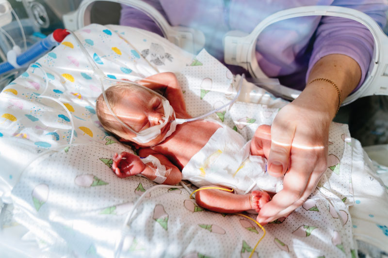
Research at Children’s and UAB shows that keeping premature infants on CPAP longer may improve lung growth.

Research at Children’s of Alabama and the University of Alabama at Birmingham suggests that keeping premature babies on nasal continuous positive airway pressure (CPAP) longer than currently practiced could significantly improve respiratory outcomes, potentially changing how neonatologists approach respiratory care for the smallest patients.

Early use of CPAP is standard for preterm infants unable to breathe on their own. It helps avoid invasive ventilation and minimizes the risk of lung injury while also increasing lung volume, which could stimulate lung growth and development.

Zaki Yazdi, M.D.

Children’s neonatologist Zaki Yazdi, M.D., conducted a pilot study as part of his fellowship to see whether extending CPAP beyond traditional stopping points benefited premature infants. Yazdi’s study, published in Archives of Disease in Childhood: Fetal & Neonatal, showed that continuing CPAP reduced episodes of drops in heart rate and oxygen levels in preterm infants. These positive findings align with groundbreaking research published this year in the American Journal of Critical Care Medicine, suggesting that extending CPAP promotes lung growth in babies born prematurely.

“We know CPAP helps premature babies with respiratory distress syndrome and apnea of prematurity,” Yazdi said. “The question we were trying to answer was: When is the best time to stop CPAP? We hypothesized that if you were to continue CPAP for a 24-hour period instead of going down to nasal cannula, you would have fewer drops in your oxygen level.”

Yazdi and the Children’s neonatology team, including neonatologist Colm P. Travers, M.D., randomized 36 infants born before 34 weeks gestation to either stop CPAP when they met Children’s traditional criteria (minimal oxygen support and few apnea episodes) or continue for an additional 24 hours. The primary outcomes were oxygen levels and other vital sign changes.

Colm Travers, M.D.

Babies who remained on CPAP an additional 24 hours experienced significantly fewer episodes of intermittent hypoxemia—defined as oxygen saturation below 85% for 10 seconds or longer—compared to those transitioned to low-flow nasal cannula. The CPAP group also had fewer heart rate drops and spent less time with low oxygen saturations.

“Even though all the markers we traditionally look at say this baby should be ready to come off CPAP, perhaps there are some more subtle things that we wouldn’t normally pick up on that suggest staying on CPAP could be helpful,” Yazdi said.

Extended CPAP isn’t without risks. Prolonged treatment can delay oral feeding, since many hospitals avoid feeding babies while on CPAP. There’s also risk of nasal breakdown from the CPAP mask interface, increased costs, and potential complications like feeding intolerance from swallowing air. However, Yazdi’s study found no negative effects during the 24-hour extension period.

The team has now received funding from the National Institute of Child Health and Human Development’s Neonatal Research Network to perform a much larger study examining extended CPAP’s effects on lung development. The multicenter, randomized clinical trial—led by UAB—will involve approximately 860 babies. Unlike Yazdi’s 24-hour study, neonates randomized to the longer CPAP arm will remain on the support for at least two weeks or until they are 34 weeks post-menstrual age. The children will then be followed for two years to assess lung function development and long-term respiratory outcomes, making it the largest controlled trial of extended CPAP to date. 

Already, Yazdi said, he and other neonatologists have noticed “a little bit of creep” toward keeping babies on CPAP longer at Children’s and other institutions.

“I don’t think we’re ready to say that this is definitely the best way to go yet,” Travers said. “But preliminary data that’s very promising suggests we need to do this larger trial to see if there’s any long-term benefit.”

“This could redefine what the standard of care could be,” Yazdi said.

Neonatology

Gut fungi can predict BPD, study shows

Research shows that the gut composition of fungi in the second week of life predicts the later development of BPD.

By Jeff Hansen (UAB)

Extremely preterm newborns who weigh less than 3.3 pounds have immature lungs that often require high levels of ventilation oxygen in the hospital. This contributes to the chronic lung disease bronchopulmonary dysplasia, or BPD, the most common cause of death for these tiny infants. BPD exacts a devastating toll on the immature lung.

In one of the most extensive studies of the microorganisms in the intestines of very preterm infants, University of Alabama at Birmingham (UAB) and University of Tennessee Health Science Center researchers show that the gut composition of fungi in the second week of life predicts the later development of BPD, weeks to months before diagnosis of that disease. They analyzed gut fungi in the first true non-meconium stool produced before two weeks of life and found that the fungal intestinal microbiome—known as the mycobiome—of infants who later developed BPD differed in community diversity, composition and interconnectivity from the infants who never got BPD, as measured by the most up-to-date bioinformatic techniques. The researchers did not find significant differences in the bacterial microbiome in those first true stools.

To show causality, researchers transferred samples of the first true stool that predicts BPD or the first true stool of newborns who did not get BPD into female mice to give them a pseudo-humanized gut microflora. In a mouse model of BPD, newborn pups from those BPD dams showed an increased in the severity of lung injury compared with newborn pups from the no-BPD dams. In loss-of-function experiments, when the female mice with the BPD-predictive stool transplant were treated with an antifungal agent before birth, that inhibition of perinatal fungal colonization reduced lung injury in the newborn pups. In contrast, a gain-of-function experiment, where the perinatal fungal colonization of dams was augmented with a species of Candida fungus common in mice, amplified BPD severity in the newborn pups.

Kent Willis, M.D.

“These findings demonstrate that features of the initial intestinal fungal microbiome are associated with the later development of BPD in premature neonates and exert a microbiome-driven effect that is transferable and modifiable in mouse models,” said Children’s of Alabama neonatologist Kent Willis, M.D., who’s also an assistant professor in the UAB Department of Pediatrics Division of Neonatology. “This suggests causality, and it suggests that the gut fungi may represent a therapeutic target in newborn lung disease.”

Willis and Ajay J. Talati, M.D., University of Tennessee Health Science Center, Memphis, Tennessee, co-led the study, published in Microbiome.

“Collectively, our analyses demonstrate that the composition of the intestinal mycobiome of infants who did not develop BPD was more uniform,” Willis said. “In contrast, those who eventually developed BPD had more disparate mycobiomes. This suggests that a particular pattern of mycobiome development may be necessary to impart resistance to the development of BPD, and failure to do so in various ways is associated with disease development.”

The first defecations of newborn infants are meconium, composed from materials ingested while in the uterus. The first true stools in the second week, the ones analyzed by Willis and Talati, are digested milk. It is known, Willis and Talati say, that fungi in adults are vital members of the human microbiota; but compared to bacteria, their non-pathological and non-parasitic functions are still poorly understood, especially in newborns.

This prospective observational cohort study included newborn stool samples collected over six years from 2017-2020 in Memphis and 2021-2022 in Birmingham. The 64 very-preterm infants in the study who did not develop BPD had an average birthweight of 2.5 pounds, and the 38 very-preterm infants who did develop BPD had an average birthweight of 1.6 pounds. Only one of the 64 no-BPD infants in the study died, while six of the 38 BPD infants died.

Neonatology

A closer look at the effects of chorioamnionitis on premature babies

The majority of preterm births stem from chorioamnionitis. (Stock photo)

The vast majority of preterm births—especially “micro-preemies” born at 22 or 23 weeks’ gestation—stem from a single cause: chorioamnionitis, an inflammation of the placenta and membranes surrounding the fetus. But Children’s of Alabama neonatologist Viral Jain, M.D., is on a mission to determine why this insidious condition occurs, the ways it affects babies’ health, and how to stop it.

Occurring in an estimated 1% to 5% of births in the United States, chorioamnionitis—often shortened to chorio—can be hard to spot. It’s typically diagnosed using clinical signs of inflammation such as fever or elevated heart rates in either the mother or the baby. But chorio often eludes clinical diagnosis, silently causing damage to the placenta and triggering preterm birth, says Jain, also an assistant professor in the Division of Neonatology at the University of Alabama at Birmingham (UAB).

Viral Jain, M.D.

“It’s a huge reason why neonatology exists, as such,” he explained. “It’s the body’s reaction when there’s inflammation to deliver the baby preterm, and all the complications that come with a preterm baby are due to chorio. In addition, the inflammation also causes direct damage to the developing organs of the baby.”

Some of the extensive research conducted on chorio has focused on its causes, which may include infection, environmental chemicals, smoking and bleeding. But scientists still have a poor understanding of why it happens, Jain notes, as well as how to catch it early enough to stop premature delivery.

Much of Jain’s research has delved into chorio’s potential health implications for babies once they’re born—and the effects can be devastating. One of his studies shows that the incidence of cerebral palsy is far higher in infants born when chorio progresses to such a severe extent it becomes funisitis, or inflammation of the umbilical cord. Jain’s findings have been somewhat controversial, he acknowledges, since cerebral palsy is already known to affect more preterm infants than those born after full-term pregnancies.

“We chose the most severe chorio babies for the study to clearly show that it affects cerebral palsy development,” Jain said. “We found that it’s about 50-50—so half the risk of cerebral palsy was from being born pre-term due to chorio, and half was the direct injury coming from inflammation to the developing brain.”

To help predict the cerebral palsy risk of these infants while they’re still in the neonatal intensive care unit (NICU)—when early intervention can more easily be planned—Jain’s research has also used MRI to look for specific markers in the brain suggesting a high risk of the disabling condition.

“We showed that chorioamnionitis insult, which started at birth, continues in these babies and that we can see those changes in the MRI and that they lead to cerebral palsy,” he said. “This means you can start early intervention on those babies to capture or reduce some of the damage.”

Another of Jain’s studies suggests that infants born early due to chorio have chronic lung damage. “It creates an immune cell dysfunction in the lung that there is continuous damage happening,” he explained. In addition to requiring longer ventilator and oxygen treatment, these babies “end up developing what we call BPD, or bronchopulmonary dysplasia, which is neonatal chronic lung disease.”

Ultimately, Jain says, his research—which has been funded by the American Heart Association and National Institutes of Health—seeks to learn how chorio propagates so doctors can impede its damage.

“The goal is to find out what treatment we can give so when it’s just mild we can stop the progression and it won’t become full-blown chorio and end up delivering the baby preterm,” he said. “If we can do that, we can prevent a lot of organ damage to the lung or brain.”  

For more information on Jain’s work on chorio, listen to this episode of the Children’s of Alabama PedsCast podcast.